Disease Images
Disease Images
Disease Images: Toxoplasmosis
Additional resources for Toxoplasmosis
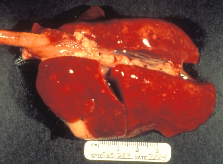
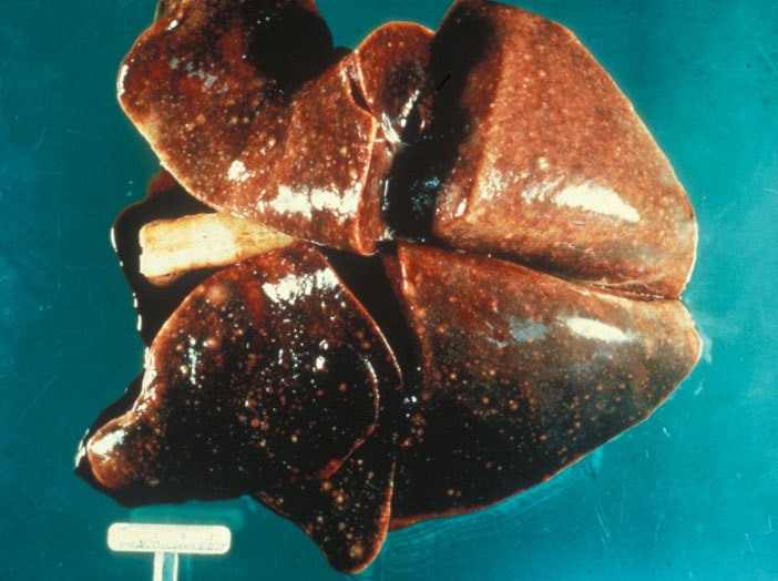
Description:
Cat, lungs. All lung lobes are noncollapsed, reddened and contain disseminated pinpoint to 2 mm diameter pale foci (interstitial pneumonia).
Photo ID: TXP_001
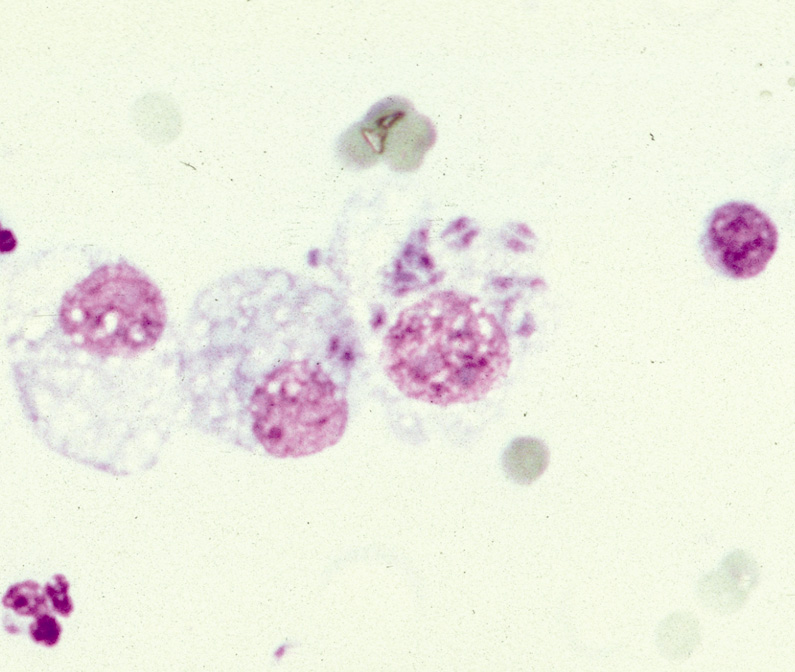
Description:
Cat, transtracheal aspirate. This transtracheal aspirate fluid contains macrophages with intracytoplasmic Toxoplasma gondii.
Photo ID: TXP_002
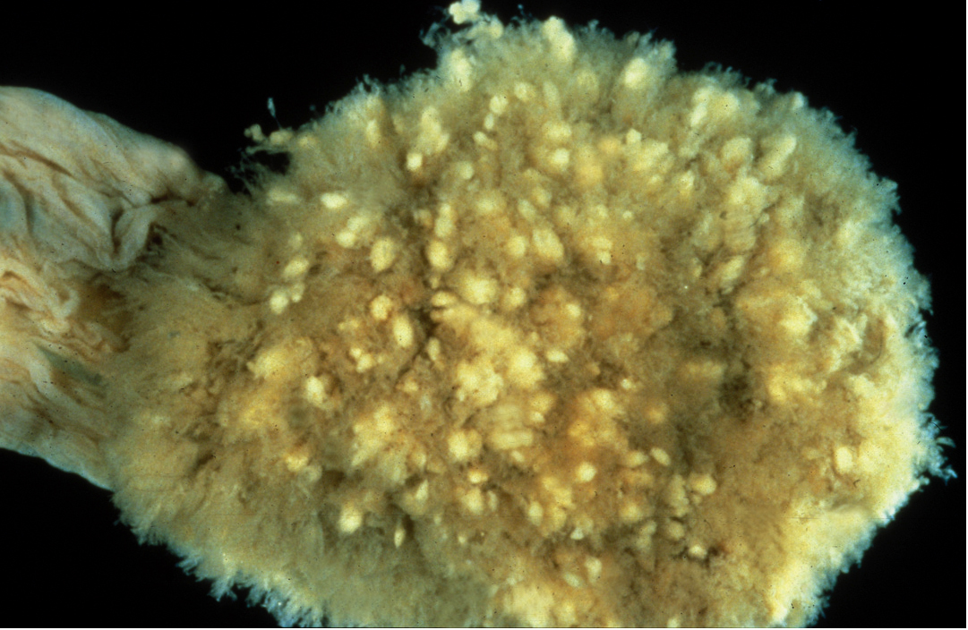
Description:
Sheep, placenta. This cotyledon has been immersed in water to demonstrate numerous pale foci of villous mineralization and necrosis.
Photo ID: TXP_003
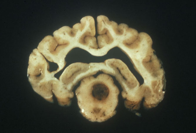
Description:
Dog, brain. Mesencephalon of the brain with a centralized brown round lesion of encephalomalacia and necrosis, and ventrally located smaller similar lesions.
Photo ID: TXP_004
Description:
Dog, lungs. Diffuse white miliary pulmonary lesions due to Toxoplasma gondii.
Photo ID: TXP_005