Disease Images
Disease Images
Disease Images: Avian Influenza
Additional resources for Avian Influenza
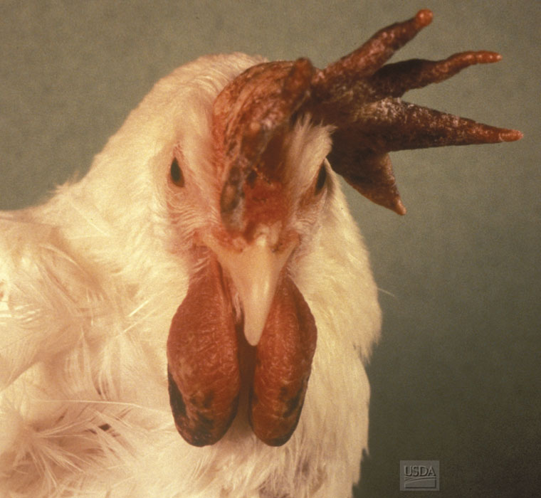
Description:
Chicken, head. The comb and wattles are congested and markedly edematous.
Photo ID: AI_001
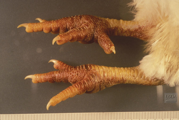
Description:
Chicken, shanks. The shanks are swollen (edema) and extensively reddened (hemorrhages).
Photo ID: AI_002

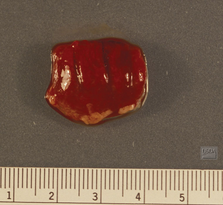
Description:
Chicken, heart. There are numerous epicardial petechiae.
Photo ID: AI_003
Description:
Chicken, lung. The lung is diffusely reddened, wet, and swollen (congestion and edema).
Photo ID: AI_004
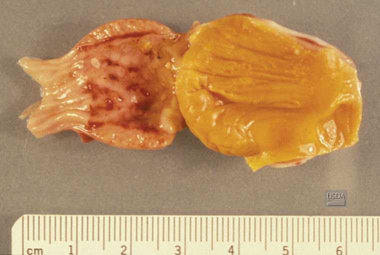
Description:
Chicken, proventriculus. There are multiple hemorrhages on the mucosal surface of the proventriculus.
Photo ID: AI_005
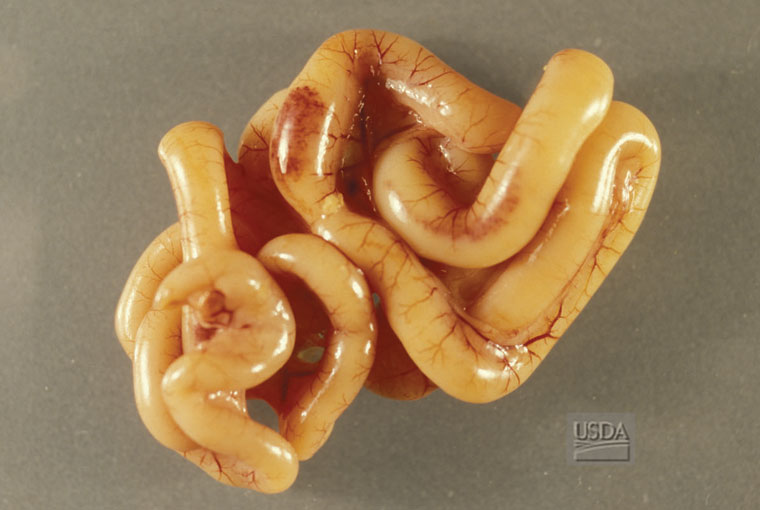
Description:
Chicken, intestine. There are serosal hemorrhages over the Peyer's patches.
Photo ID: AI_006