Disease Images

Disease Images: Trichuriasis

Additional resources for Trichuriasis


Description:
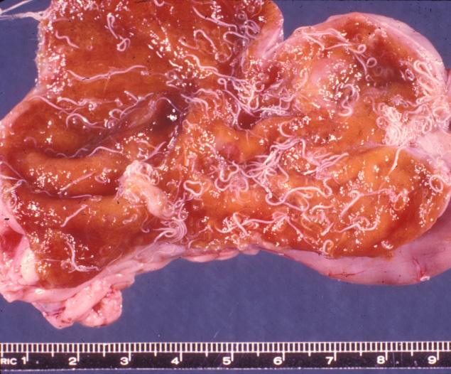
Dog, cecum. The mucosa is thickened, hyperemic, and covered by many adult Trichuris vulpis; the thicker, caudal portion of the nematode is most easily seen

Credit: AFIP
Photo ID: TRU_001

Description:
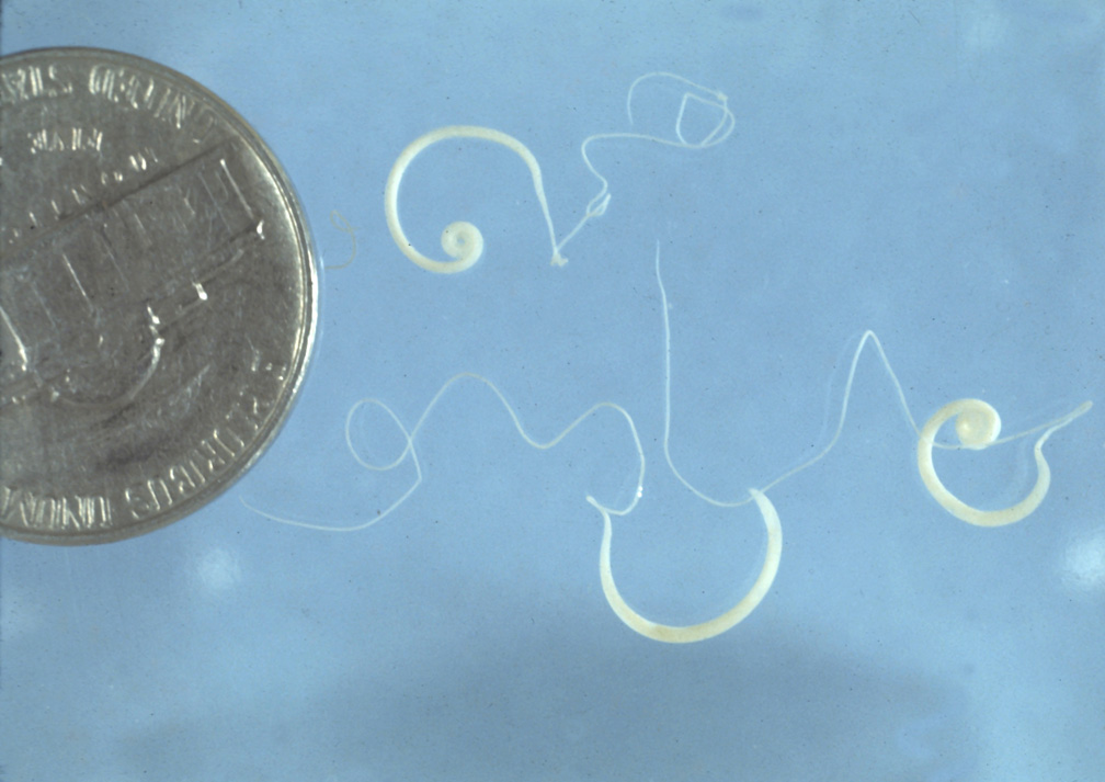
Trichuris vulpis. The cranial portion of the adult whipworm is thin and filamentous.

Credit: Dr. J. Greve, Iowa State University, College of Veterinary Medicine, Department of Veterinary Pathology
Photo ID: TRU_002

Description:
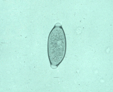
Human, feces. Whipworm eggs are oval, thick-shelled, and have transparent plugs at each pole.

Credit: AFIP
Photo ID: TRU_003

To use images and associated descriptions contained on this website, please contact the CFSPH.

These images were annotated by Dr. Steve Sorden and Dr. Claire Andreasen and funding was provided by a USDA Higher Education Challenge Grant in collaboration with the Iowa State University Department of Veterinary Pathology, Center for Food Security and Public Health (CFSPH), Armed Forces Institute of Pathology (AFIP), and Plum Island Animal Disease Center (PIADC).