Disease Images
Disease Images
Disease Images: Rift Valley Fever
Additional resources for Rift Valley Fever
Description:
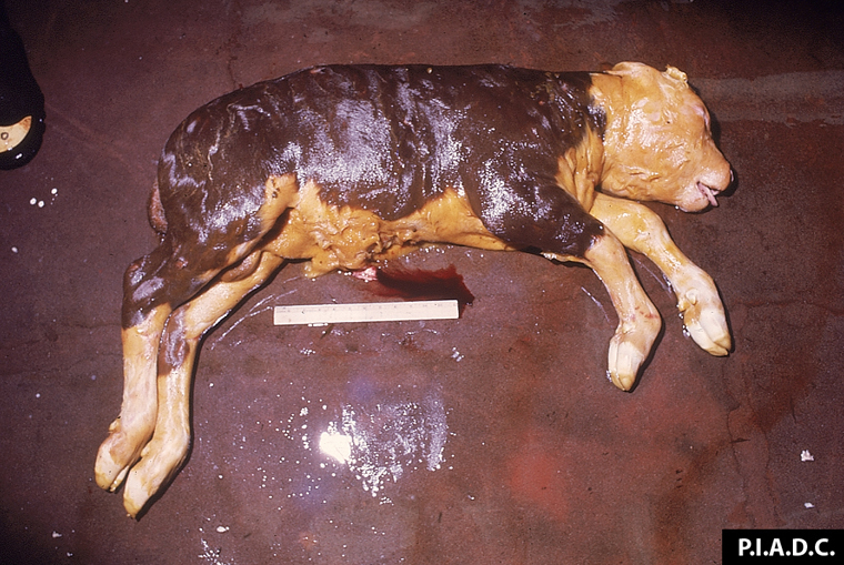
Bovine, fetus. The skin of this emphysematous fetus is stained with meconium.
Photo ID: RVF_001
Description:
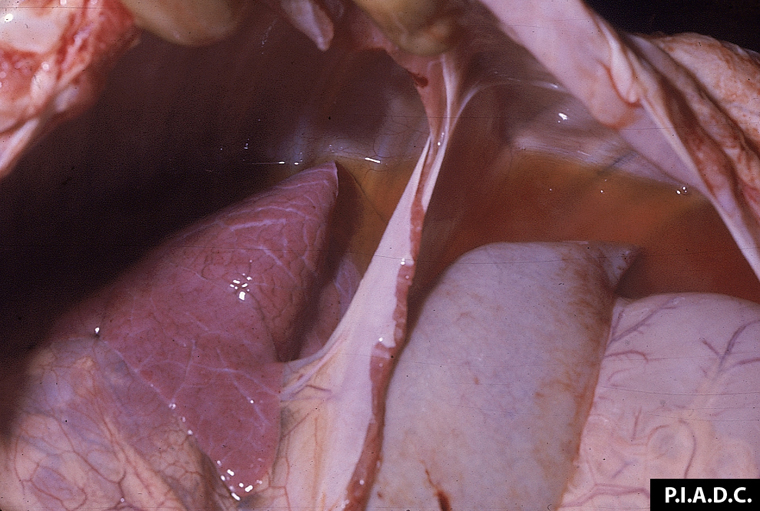
Sheep, fetus. Both the pleural and peritoneal cavities contain excessive clear, straw-colored fluid.
Photo ID: RVF_002
Description:
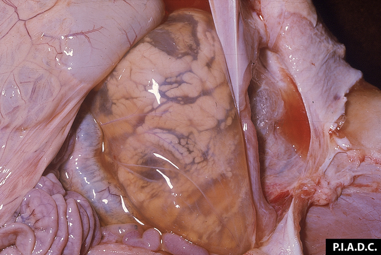
Sheep, fetus, kidney. There is severe perirenal edema.
Photo ID: RVF_003
Description:
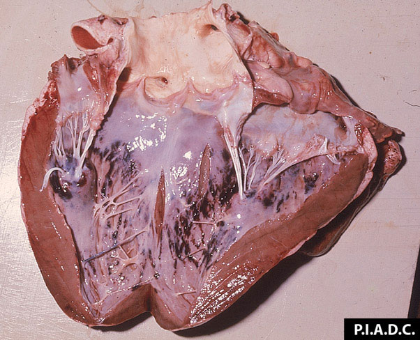
Sheep, heart. The ventricular endocardium contains many hemorrhages.
Photo ID: RVF_004
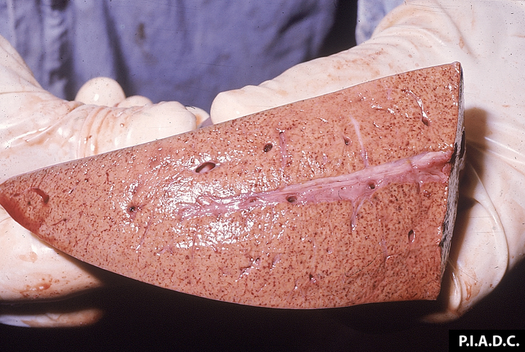
Description:
Sheep, liver. The cut surface of the swollen liver is pale and contains many petechiae.
Photo ID: RVF_005
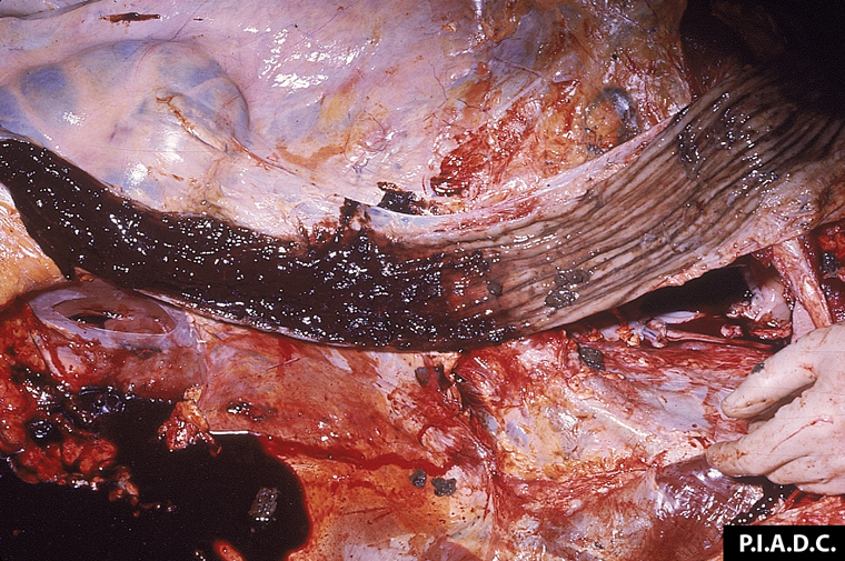
Description:
Sheep, colon. Severe hemorrhagic colitis.
Photo ID: RVF_006
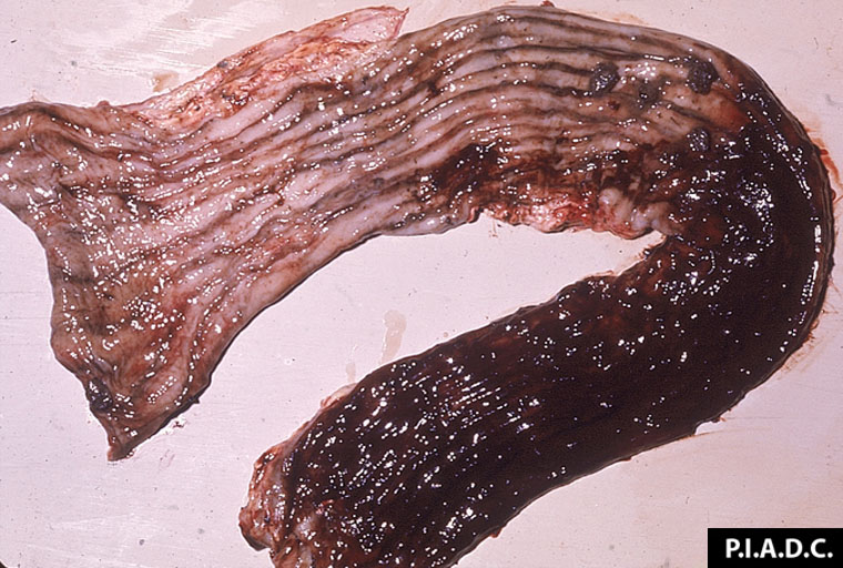
Description:
Sheep, colon. There is severe locally extensive mucosal hemorrhage.
Photo ID: RVF_007
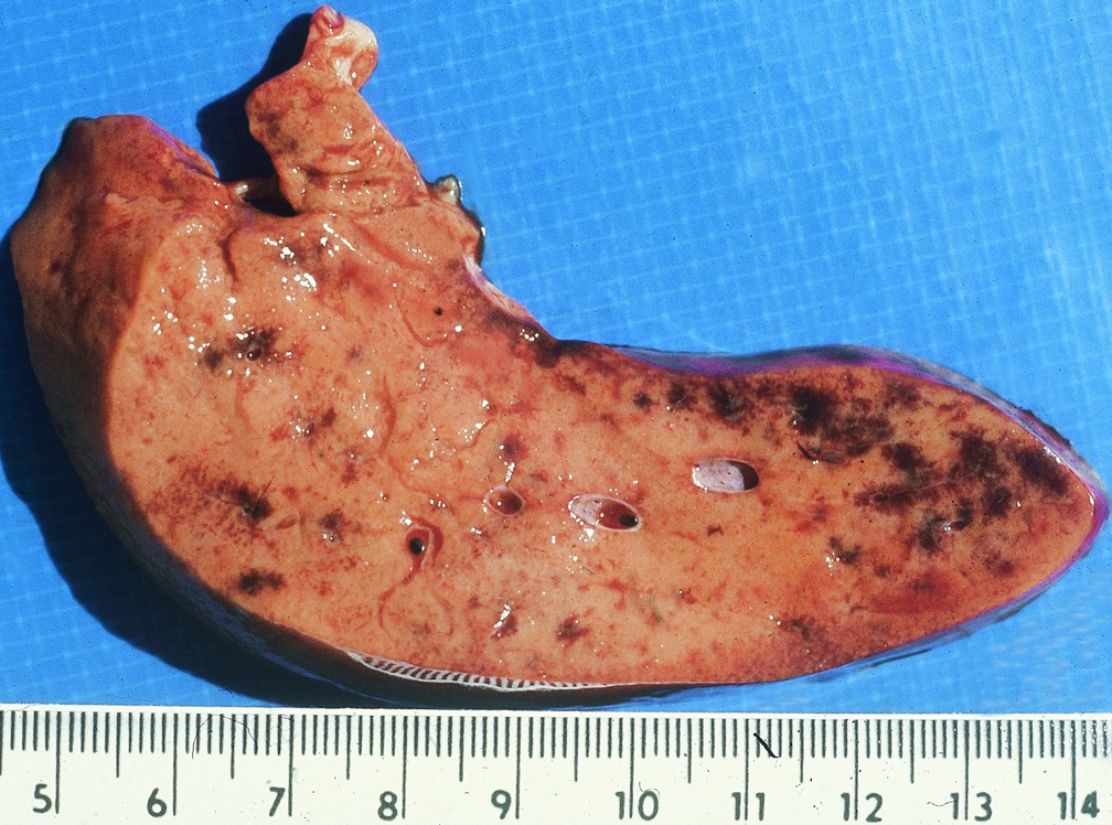
Description:
Sheep, liver. Section reveals that the liver is pale, swollen and contains multiple foci of hemorrhage.
Photo ID: RVF_008
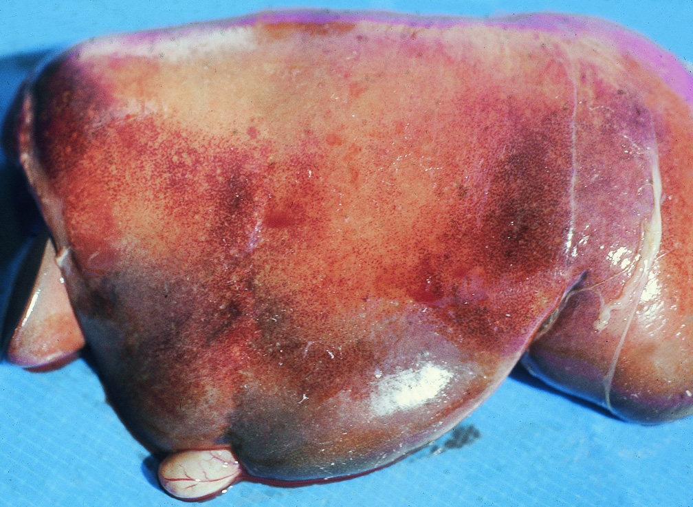
Description:
Sheep, liver. Liver is pale and swollen and contains many areas of severe congestion.
Photo ID: RVF_009