Disease Images
Disease Images
Disease Images: Malignant Catarrhal Fever
Additional resources for Malignant Catarrhal Fever
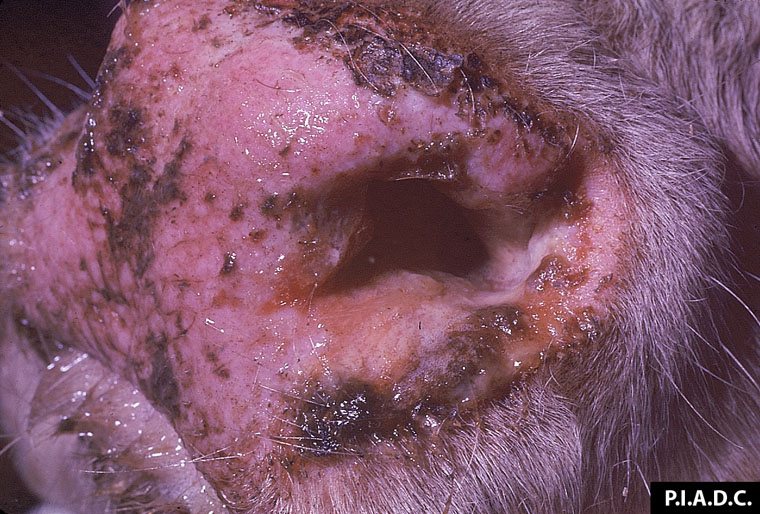
Description:
Bovine, muzzle. Multiple shallow erosions are filled with dried nasal exudate.
Photo ID: MCF_001
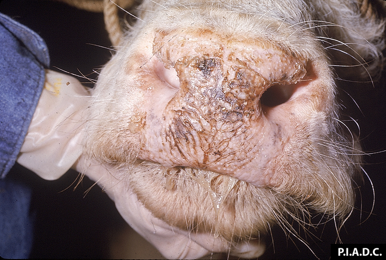
Description:
Bovine, muzzle. The muzzle is hyperemic, multifocally covered by adherent mucopurulent exudate, and contains many shallow erosions.
Photo ID: MCF_002
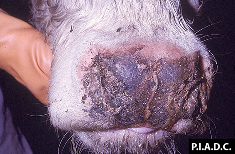
Description:
Bovine, muzzle. There is diffuse superficial necrosis of the muzzle.
Photo ID: MCF_003
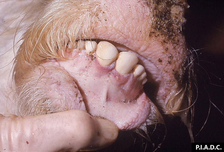
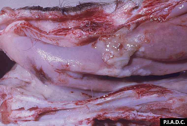
Description:
Bovine, oral mucosa. There is a gingival hyperemia and focal erosion.
Photo ID: MCF_004
Description:
Bovine, hard palate. There are multiple coalescing mucosal erosions.
Photo ID: MCF_005
Description:
Bovine, skin. There are numerous raised plaques (multifocal dermatitis).
Photo ID: MCF_006
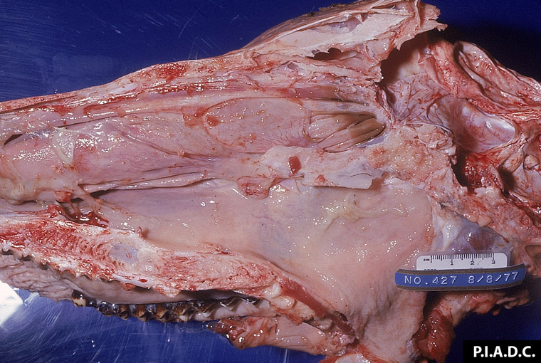
Description:
Bovine, head, sagittal section. Mucoid exudate multifocally covers the nasal and pharyngeal mucosa.
Photo ID: MCF_007
Description:
Bovine, nasal turbinate. There is a small amount of mucoid exudate.
Photo ID: MCF_008
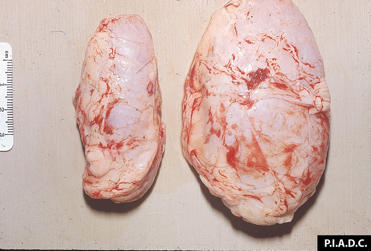
Description:
Bovine, prescapular lymph nodes: Moderately (left) to markedly enlarged (right) due to MCF.
Photo ID: MCF_009
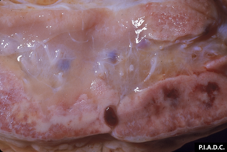
Description:
Bovine, prescapular lymph node. There are foci of hemorrhage (and necrosis) in the cortex, and the medulla is edematous.
Photo ID: MCF_010
Description:
Bovine, prescapular lymph node. There are foci of hemorrhage (and necrosis) in the cortex, and the medulla is edematous.
Photo ID: MCF_011
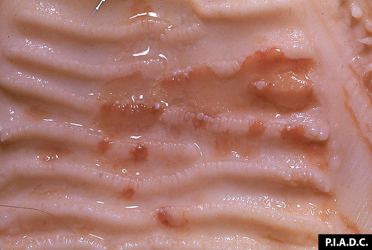
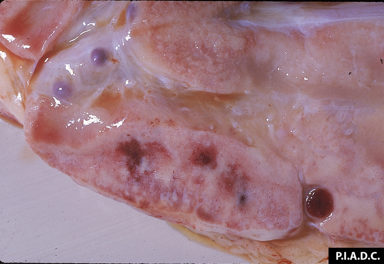
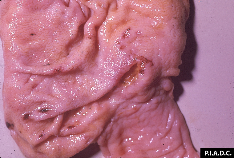
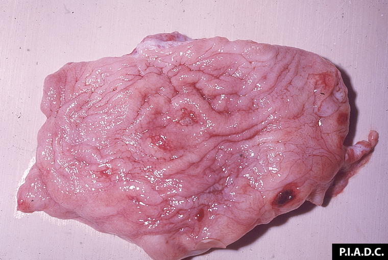
Description:
Bovine, omasum. Omasal leaves contain multiple pale foci of necrosis; on the right there are several ulcers.
Photo ID: MCF_012
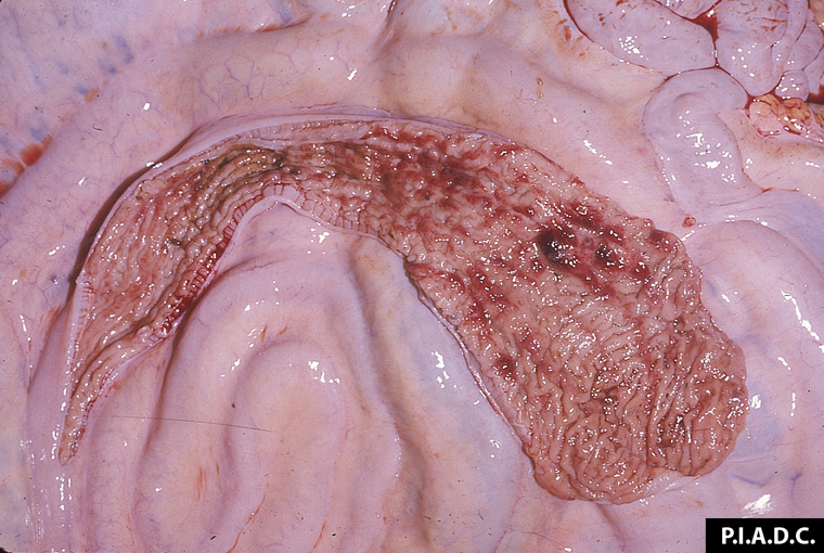
Description:
Bovine, cecum and ileum. There are scattered small foci of mucosal hemorrhage and erosion.
Photo ID: MCF_013
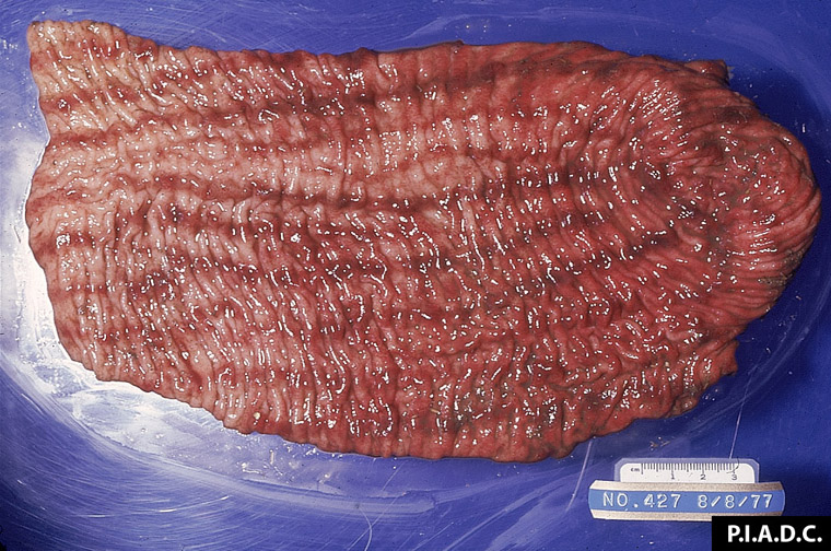
Description:
Bovine, spiral colon. There are multiple mucosal hemorrhages.
Photo ID: MCF_014
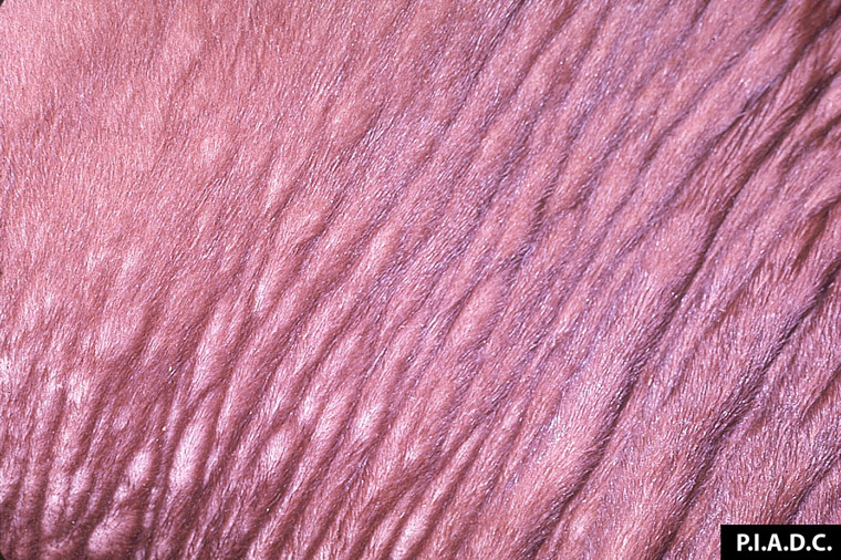
Description:
Bovine, colon. There is severe longitudinal linear congestion of the mucosa.
Photo ID: MCF_015
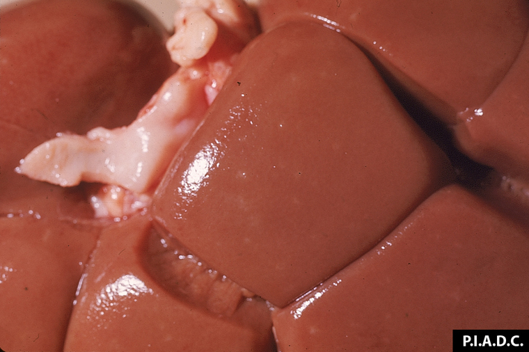
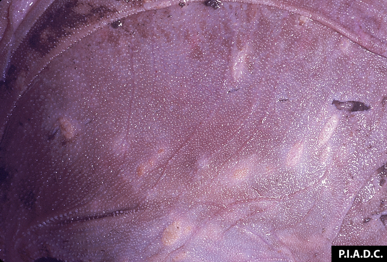
Description:
Bovine, kidney. Multiple pale foci in the cortex are foci of institial nephritis.
Photo ID: MCF_016
Description:
Bovine, urinary bladder. The mucosal surface contains several small erosions and one large hemorrhagic ulcer.
Photo ID: MCF_017