Disease Images
Disease Images
Disease Images: Maedi–Visna
Additional resources for Maedi–Visna
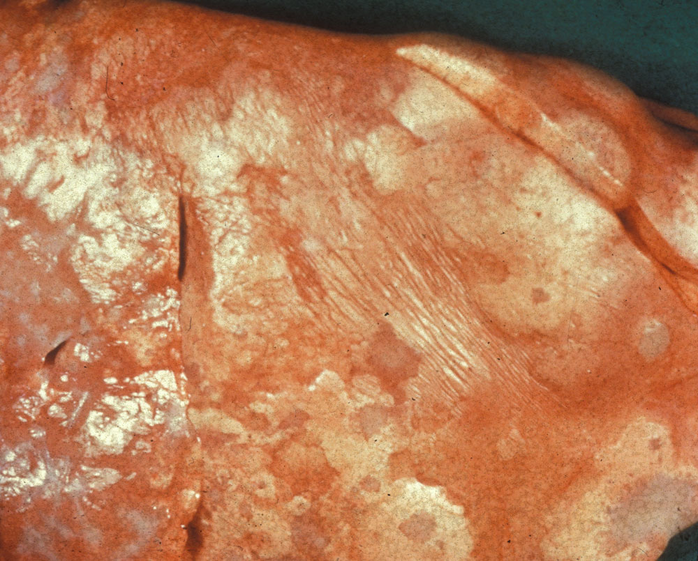
Description:
Sheep, lung. Lung fails to deflate and contains coalescing multifocal gray-white nodules/plaques (proliferative lymphocytes and pneumocytes) with adjacent atelectatic depressed parenchyma (red-pink).
Photo ID: MV_001
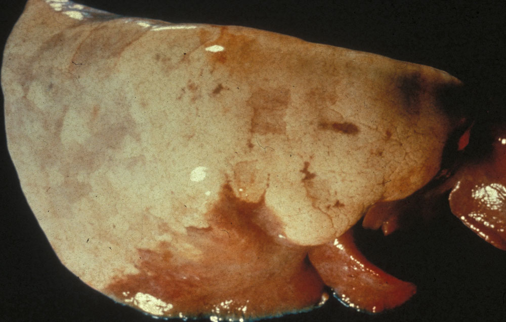
Description:
Sheep, lung. Lung fails to deflate with pale gray coalescing proliferative areas and cranioventral atelectasis (reddish area)
Photo ID: MV_002