Disease Images
Disease Images
Disease Images: Lyme Disease
Additional resources for Lyme Disease
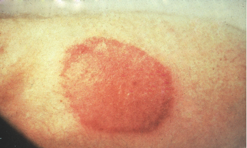
Description:
Human, extremity, skin. Erythematous macule (erythema migrans) surrounded by an intense zone of hyperemia and/or hemorrhage.
Photo ID: LYM_001
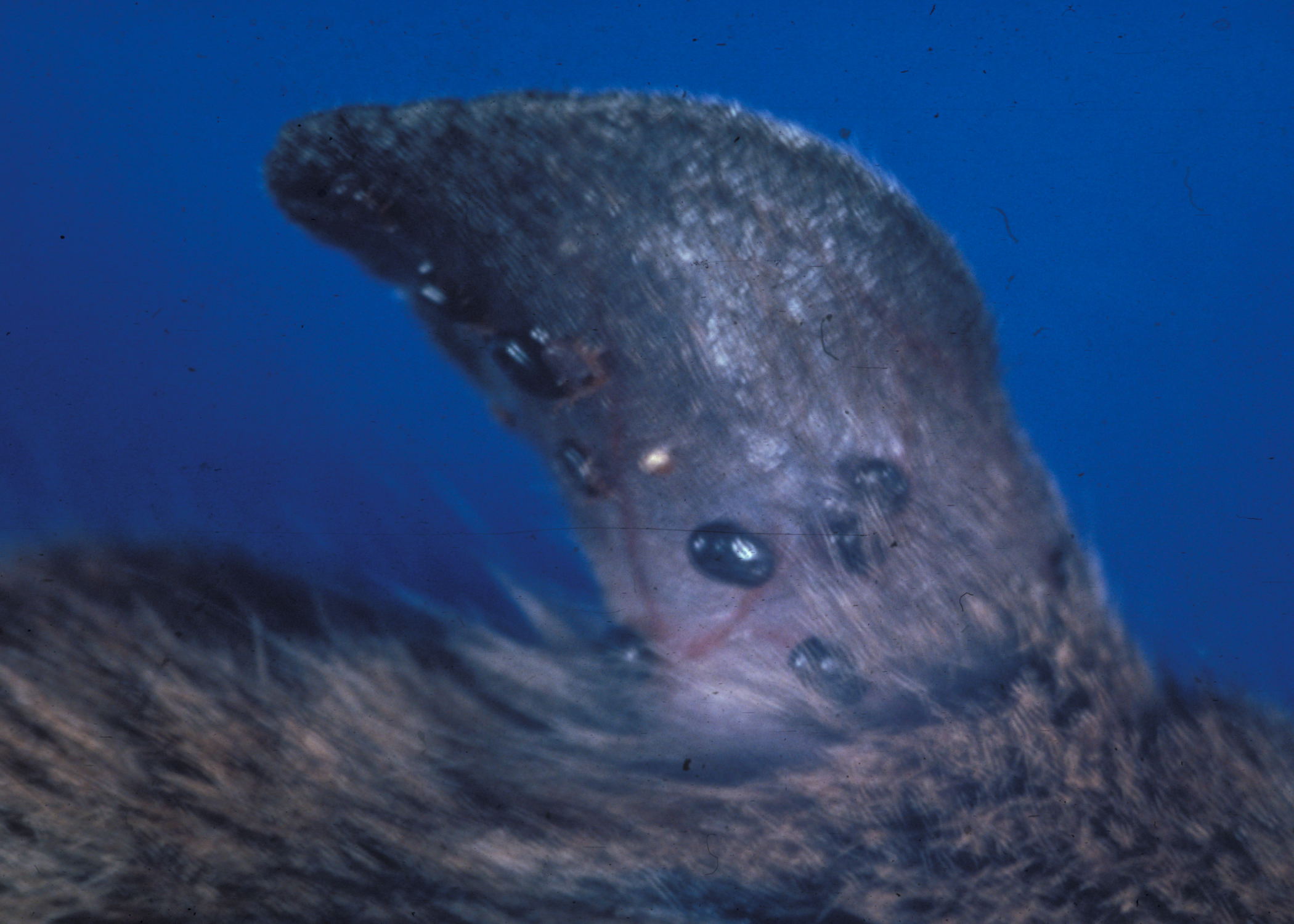
Description:
Ear, ticks. Attached Ixodes scapularis (I. dammini) with Borrelia (Lyme disease).
Photo ID: LYM_002
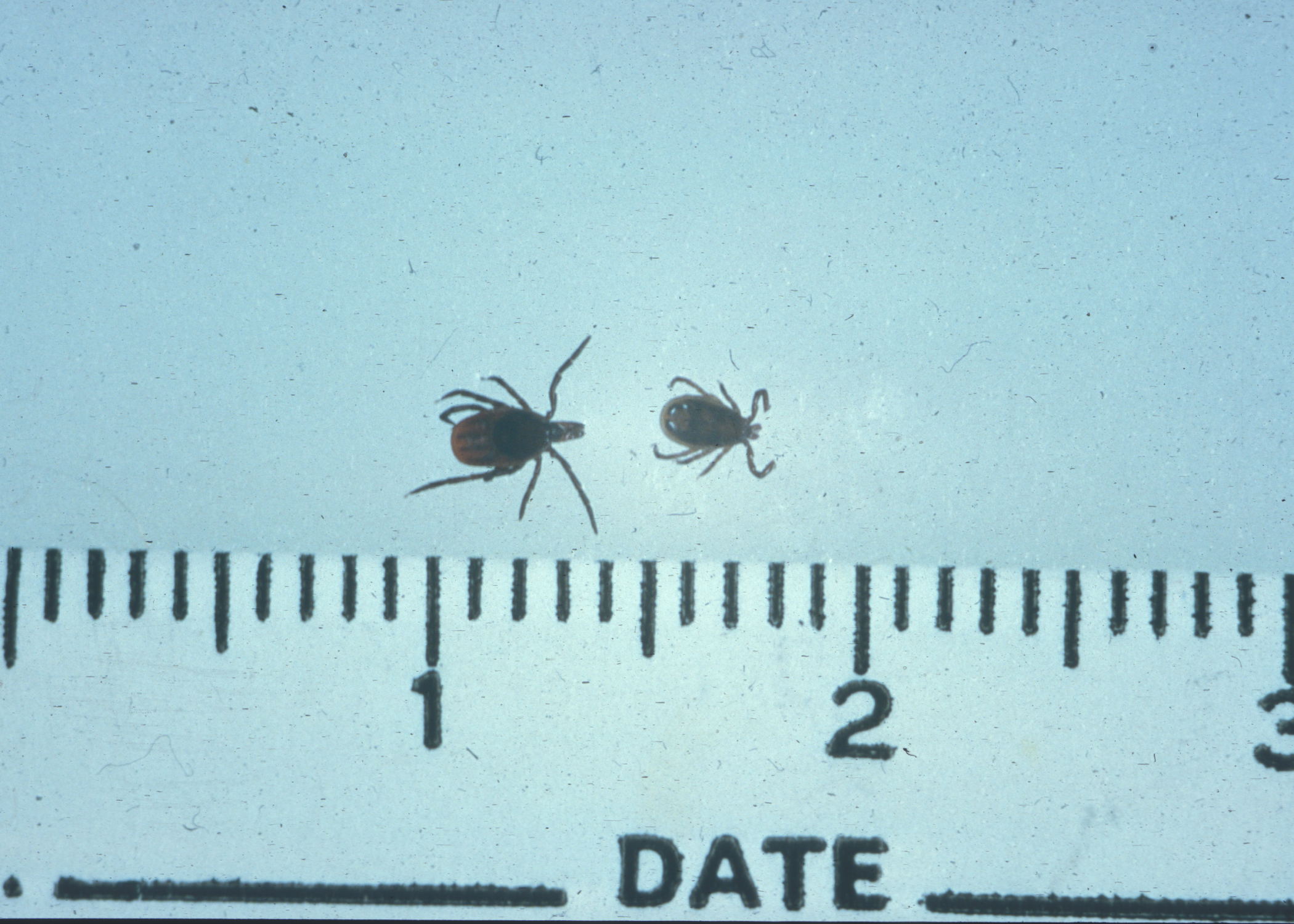
Description:
Tick. Ixodes scapularis (I. dammini) female and male carriers of Lyme disease.
Photo ID: LYM_003
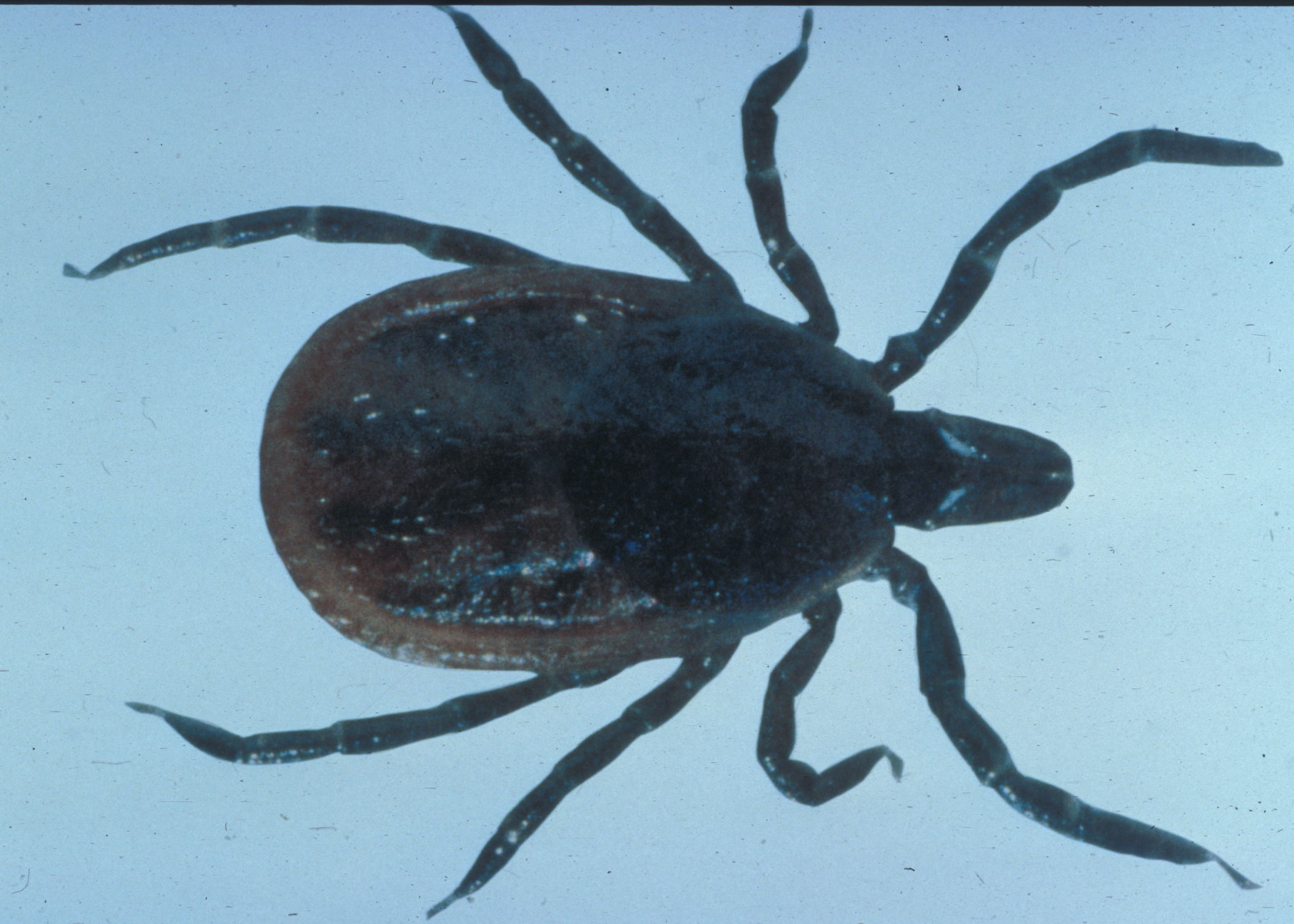
Description:
Tick. Ixodes scapularis (I. dammini) carrier tick of Lyme Disease.
Photo ID: LYM_004