Disease Images
Disease Images
Disease Images: Foot and Mouth Disease
Additional resources for Foot and Mouth Disease
Description:
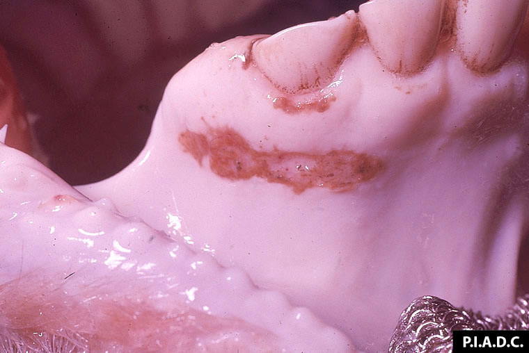
Bovine, gingiva. There is an elongate erosion (ruptured vesicle) ventral to the incisors.
Photo ID: FMD_001
Description:
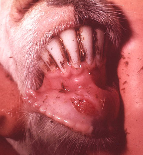
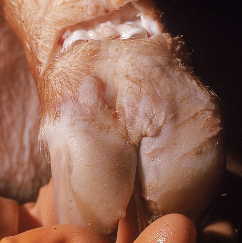
Goat, oral mucosa. There is a large erosion (ruptured vesicle) on the rostral mandibular buccal mucosa.
Photo ID: FMD_002
Description:
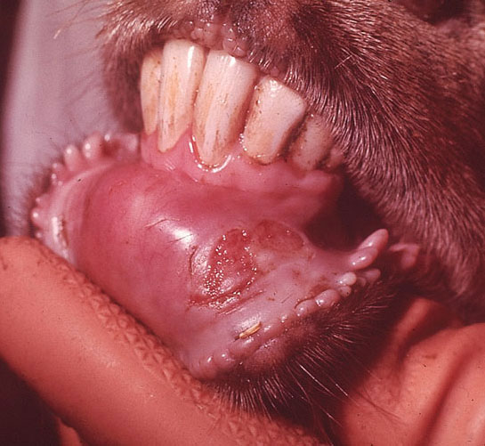
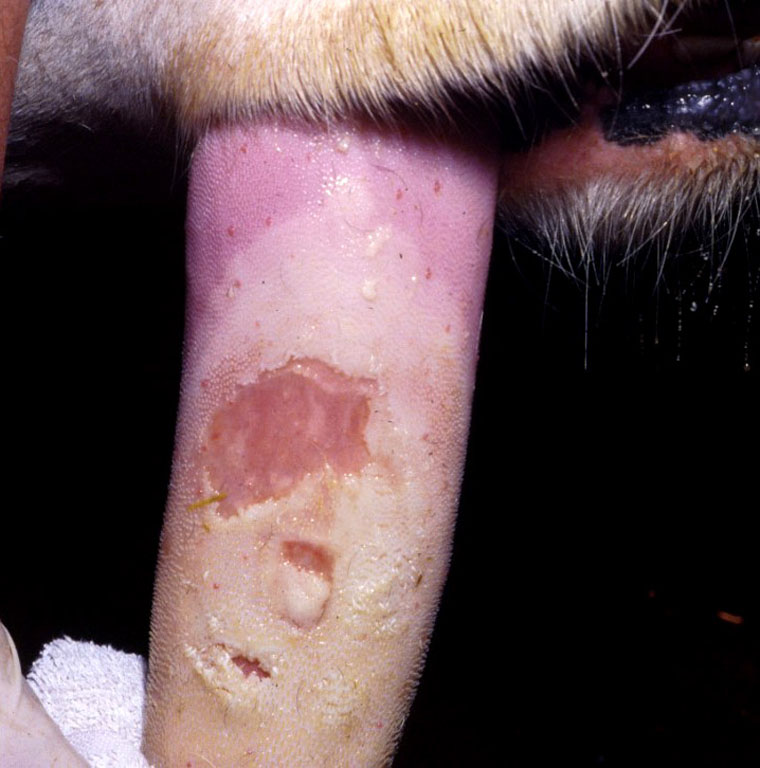
Goat, oral mucosa. There is a large, partially re-epithelialized (healing) erosion on the rostral mandibular buccal mucosa.
Photo ID: FMD_003
Description:
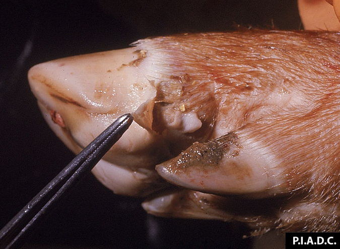
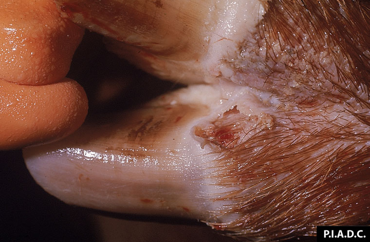
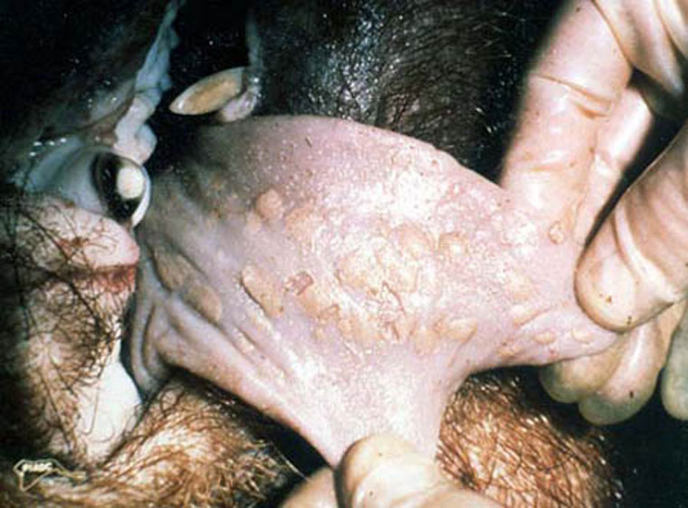
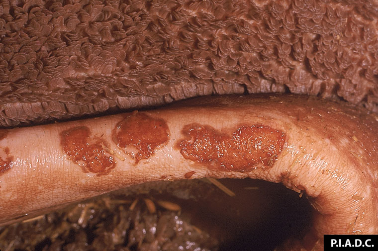
Pig, foot. There is a ruptured vesicle of the caudal-lateral coronary band, with undermining of the heel.
Photo ID: FMD_004
Description:
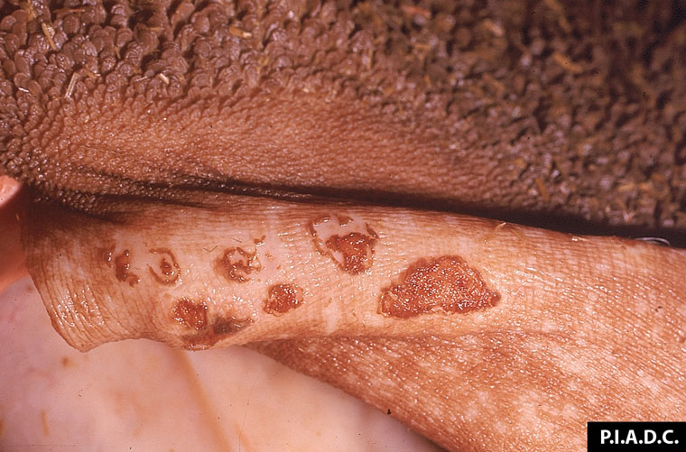
Pig, foot. A ruptured vesicle of the coronary band extends into the interdigital skin.
Photo ID: FMD_005
Description:
Pig, foot. There is an intact vesicle on the caudal coronary band of the left claw, and a cleft (ruptured vesicle) on the heel bulb of the right claw.
Photo ID: FMD_006
Description:
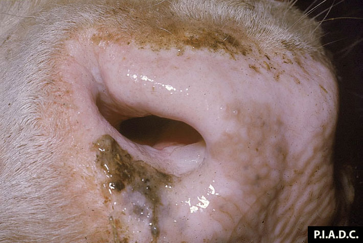
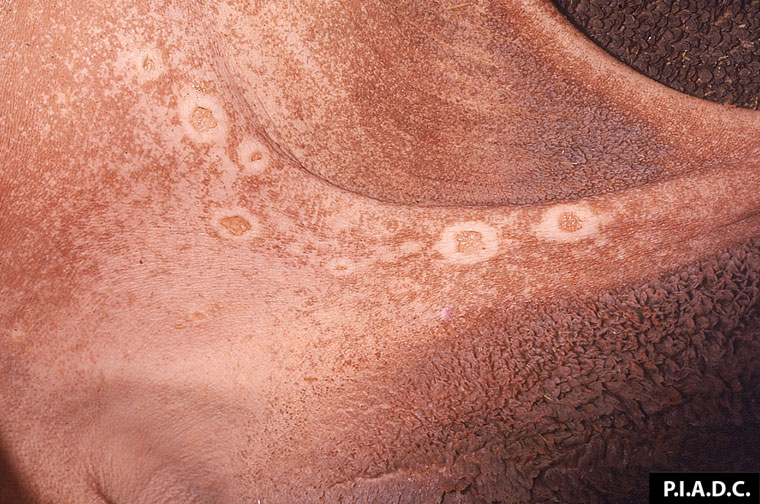
Bovine, muzzle. Within the naris, the ventromedial mucosa contains an intact vesicle.
Photo ID: FMD_007
Description:
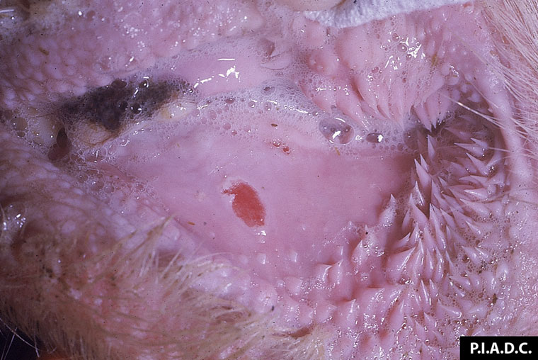
Bovine, lip. The buccal mucosa contains an erosion (ruptured vesicle).
Photo ID: FMD_008
Description:
Tongue. There are multiple large mucosal erosions
and ulcers.
Photo ID: FMD_009
Description:
Bovine, tongue. A large area of undermined epithelium (bulla) is centrally eroded; this lesion probably resulted from coalescence of several smaller lesions.
Photo ID: FMD_010

Description:
Bovine, tongue. Several healing vesicles have yellow-tan margins.
Photo ID: FMD_011
Description:
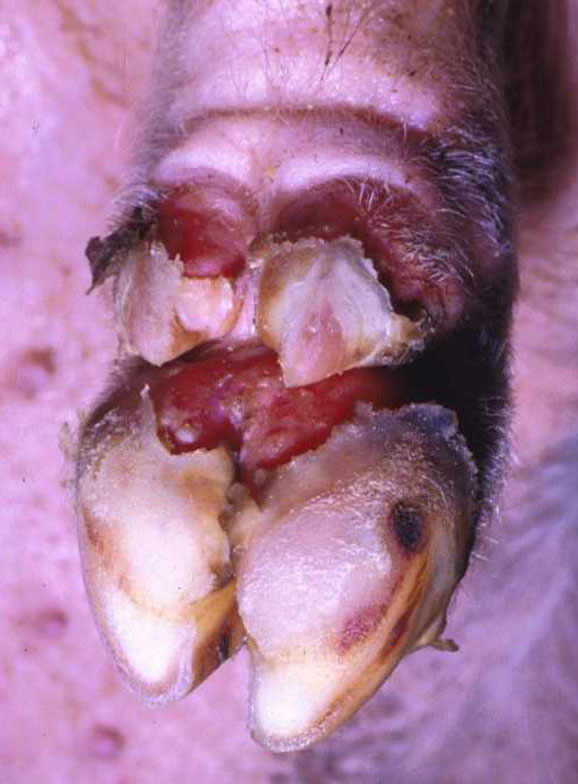
Pig, foot. Large clefts at the coronary bands precede sloughing of the claws.
Photo ID: FMD_012
Description:
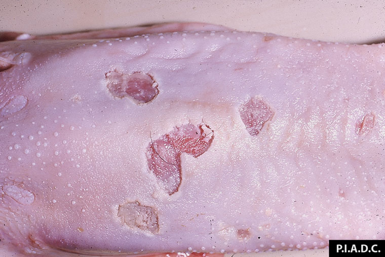
Pig, tongue. Many ("dry") vesicles are ruptured and lack fluid.
Photo ID: FMD_013
Description:
Rumen mucosa, higher magnification. There are
several irregularly shaped erosions (ruptured vesicles) on the pillar.
Photo ID: FMD_014
Description:
Rumen mucosa, dorsal sac, low magnification. There are several irregularly shaped erosions (ruptured vesicles) on the pillars. The pale margins are undermined epithelium (vesicle remnants).
Photo ID: FMD_015
Description:
Rumen mucosa, higher magnification. There are
several irregularly shaped erosions (ruptured vesicles) on the pillar.
Photo ID: FMD_016
Description:
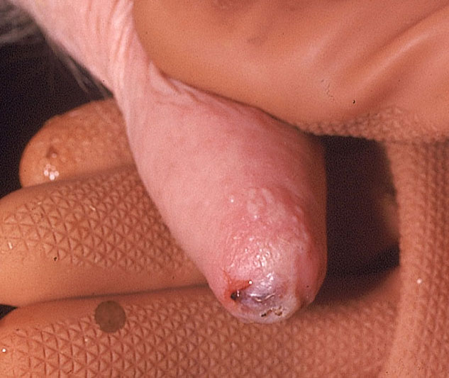
Teat. There is a ruptured vesicle on the end of the teat.
Photo ID: FMD_017
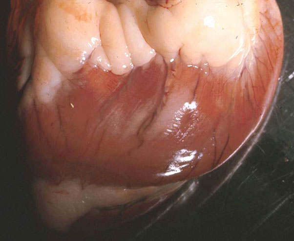
Description:
Sheep, heart. There is a pale area of myocardial necrosis visible from the epicardial surface.
Photo ID: FMD_018