Disease Images

Disease Images: Echinococcosis

Additional resources for Echinococcosis


Description:
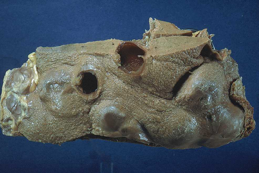
Skunk, liver. The inner surface of the cyst is lined by hydatid sand, and the cyst is surrounded by a thick capsule of fibrous connective tissue.

Credit: Iowa State University, College of Veterinary Medicine
Photo ID: ECH_001

Description:
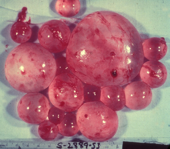
Human, liver. Multiple thin-walled hydatid cysts project from the capsular surface of the liver.

Credit: AFIP
Photo ID: ECH_002

To use images and associated descriptions contained on this website, please contact the CFSPH.

These images were annotated by Dr. Steve Sorden and Dr. Claire Andreasen and funding was provided by a USDA Higher Education Challenge Grant in collaboration with the Iowa State University Department of Veterinary Pathology, Center for Food Security and Public Health (CFSPH), Armed Forces Institute of Pathology (AFIP), and Plum Island Animal Disease Center (PIADC).