Disease Images
Disease Images
Disease Images: Cryptococcosis
Additional resources for Cryptococcosis
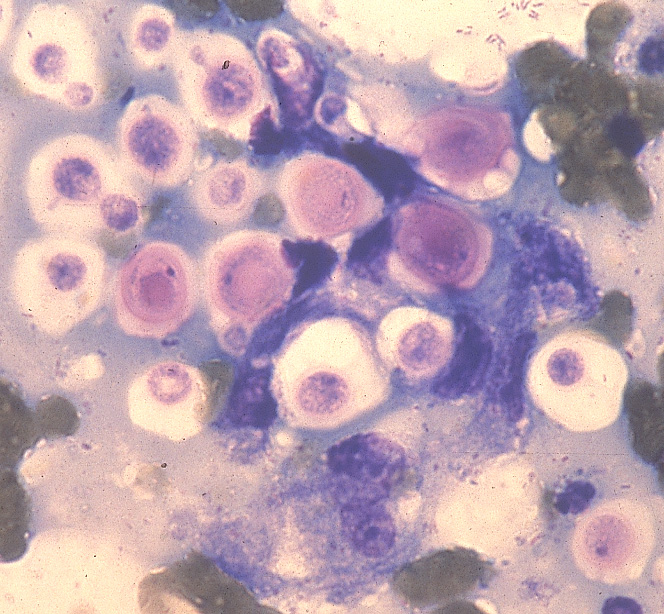
Description:
Cat. This aspirate from a cutaneous lesion contains numerous Cryptococcus neoformans yeast organisms surrounded by a nonstaining capsule. Narrow-based budding can be seen.
Photo ID: CRC_001
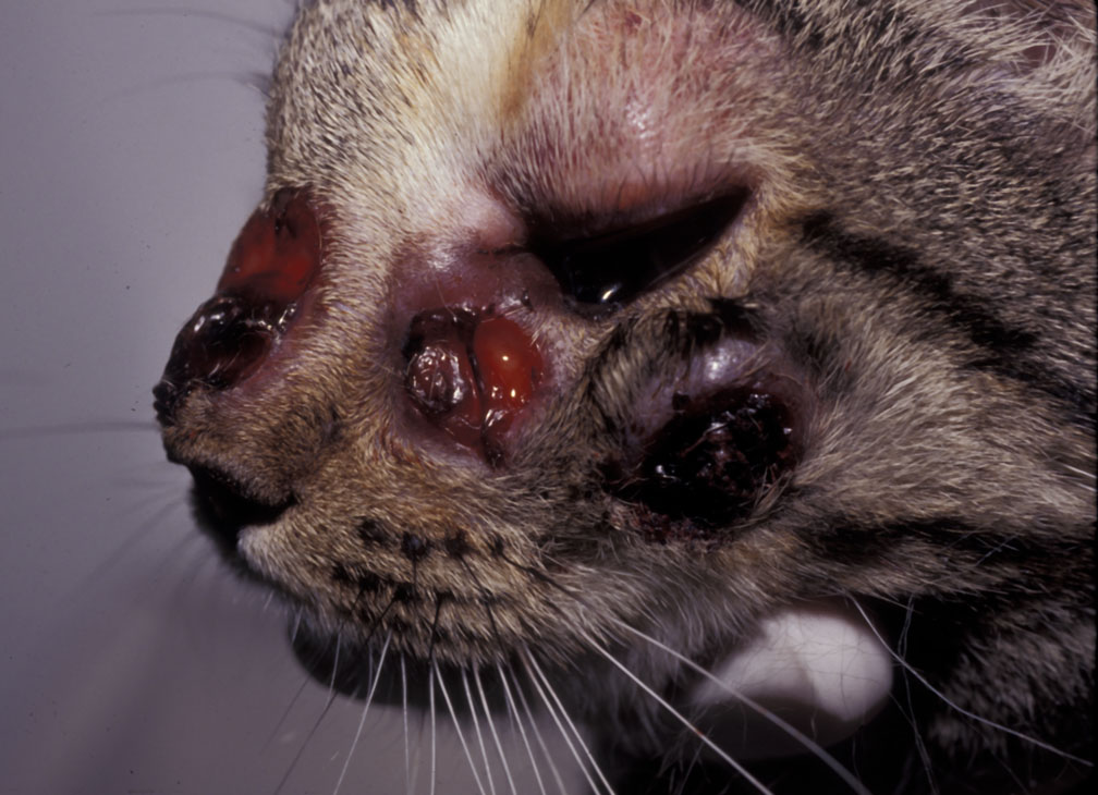
Description:
Cat. There are multiple foci of ulcerative dermatitis; the rostral lesion likely resulted from extension of cryptococcal rhinitis through the facial bones.
Photo ID: CRC_002