Disease Images
Disease Images
Disease Images: Coccidioidomycosis
Additional resources for Coccidioidomycosis
Description:
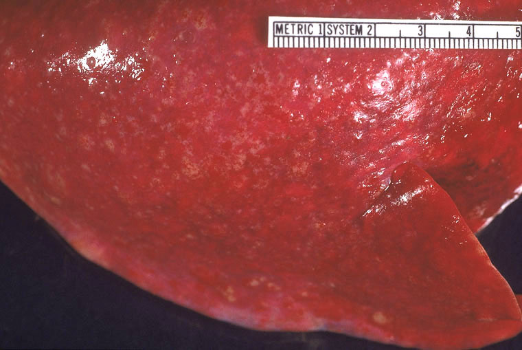
Wallaby, lung. Lung tissue is noncollapsed and contains numerous small colaescing firm pale nodules (granulomas).
Photo ID: COC_001
Description:
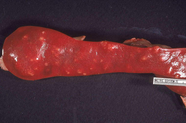
Wallaby, spleen. The spleen contains many firm pale nodules (granulomas).
Photo ID: COC_002
Description:
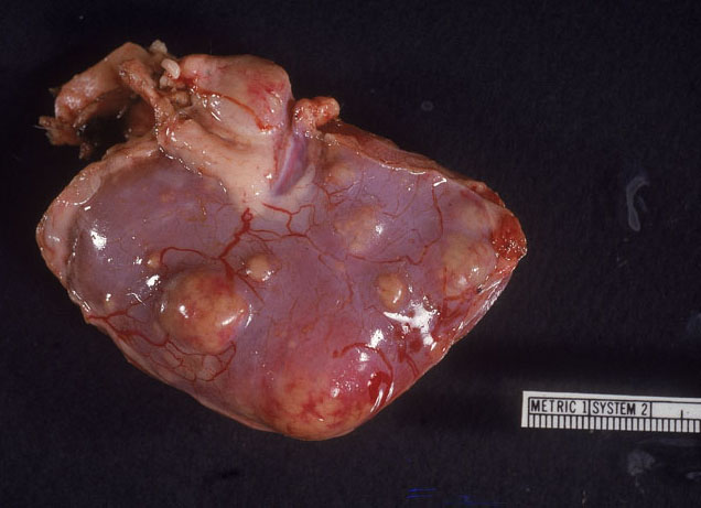
Wallaby, kidney. The kidney is distorted by multiple variably-sized firm pale raised nodules (granulomas).
Photo ID: COC_003
Description:
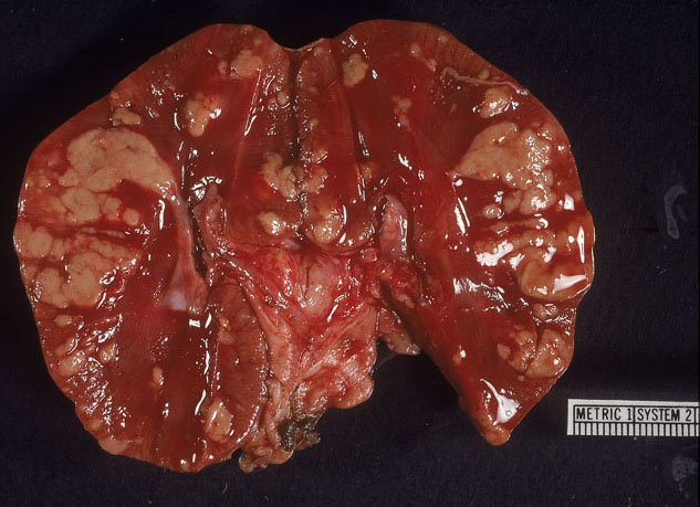
Wallaby, kidney. Section of the kidney reveals many coalescing pale raised nodules (granulomas).
Photo ID: COC_004
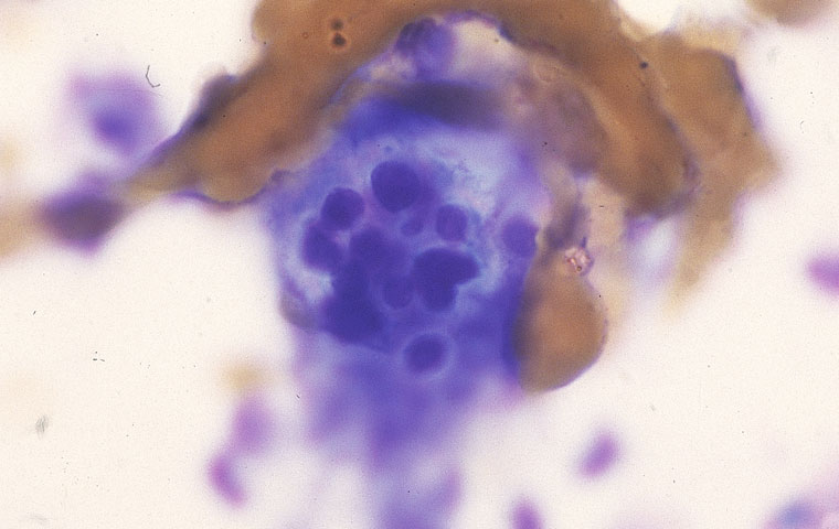
Description:
A Coccidioides immitis spherule contains multiple endospores.
Photo ID: COC_005
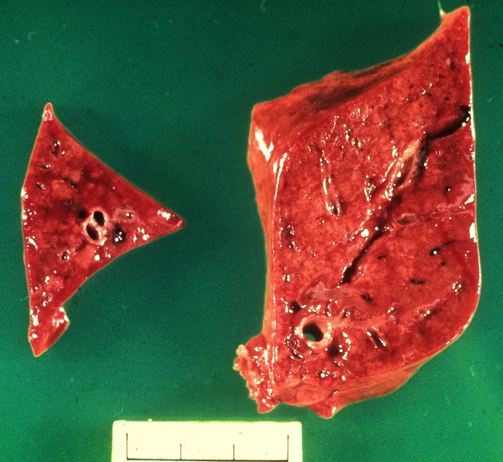
Description:
Dog, lung. Cross section of lung reveals multifocal to coalescing pale firm areas (granulomas).
Photo ID: COC_006