Disease Images
Disease Images
Disease Images: Baylisascariasis
Additional resources for Baylisascariasis
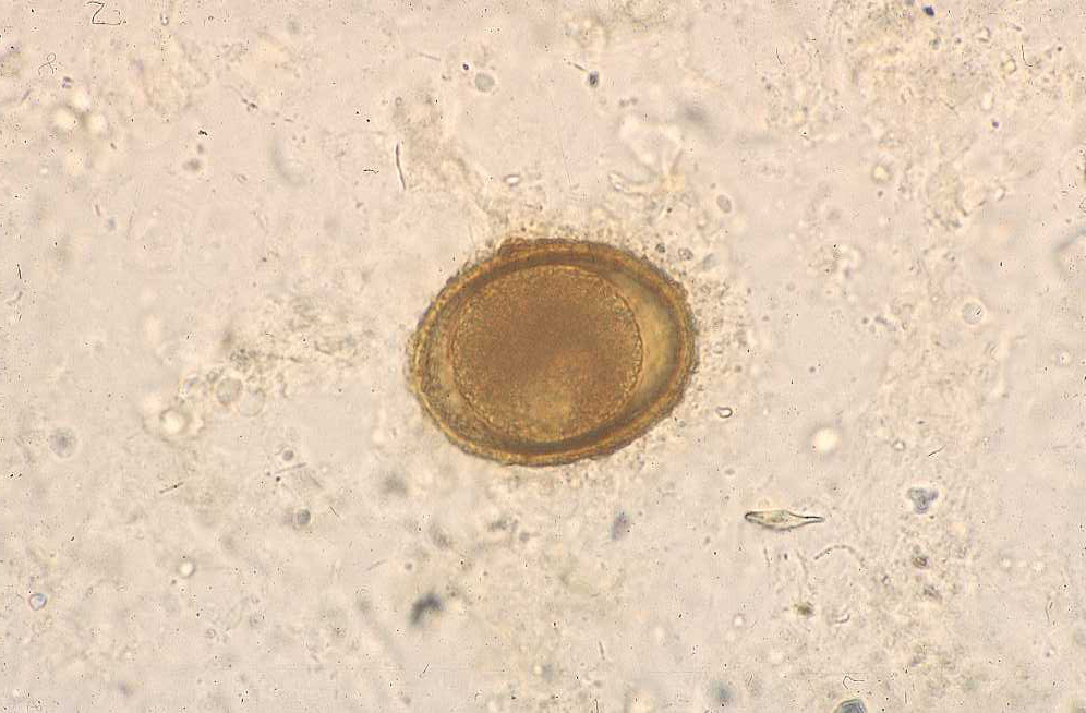
Description:
Raccoon, feces. Baylisascaris procyonis eggs are typical ascarid eggs with thick, finely pitted shells; they are slightly smaller than Toxocara canis eggs.
Photo ID: BAY_001
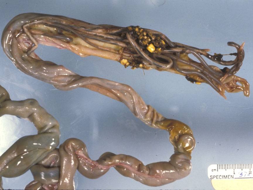
Description:
Raccoon, intestine. This partially opened small intestine contains many adult B. procyonis.
Photo ID: BAY_002