Disease Images
Disease Images
Disease Images: African Horse Sickness
Additional resources for African Horse Sickness
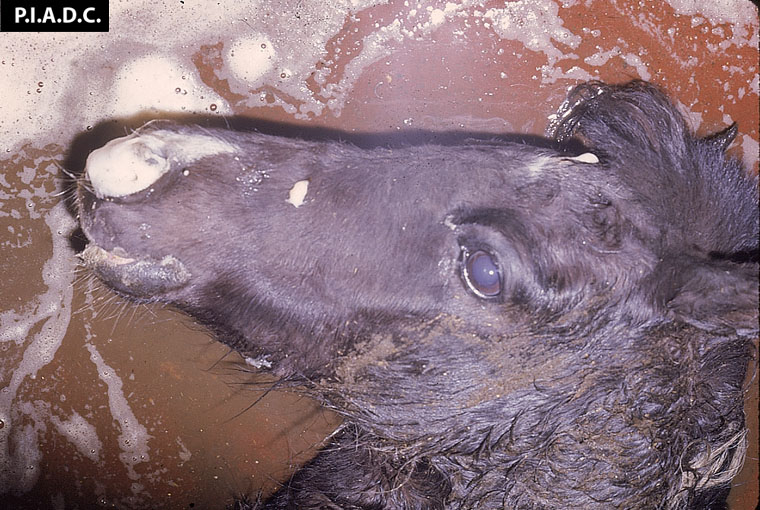
Description:
Horse. Abundant froth draining from the nostrils reflects severe pulmonary edema.
Photo ID: AHS_001
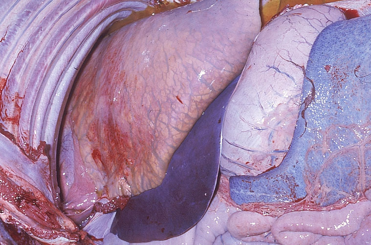
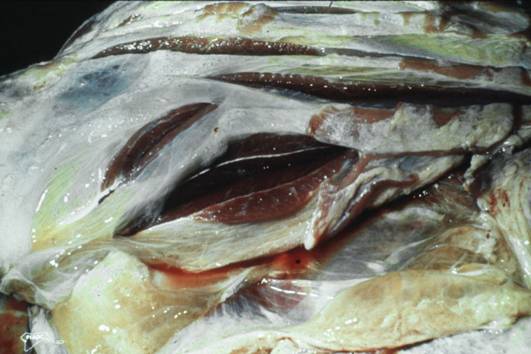
Description:
Horse. The lung exhibits severe interlobular edema. There are petechiae on the pulmonary pleura and the splenic capsule.
Photo ID: AHS_002
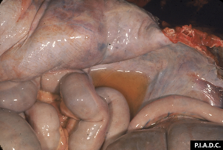
Description:
Horse, peritoneal cavity. There is excessive straw-colored fluid (hydroperitoneum).
Photo ID: AHS_003
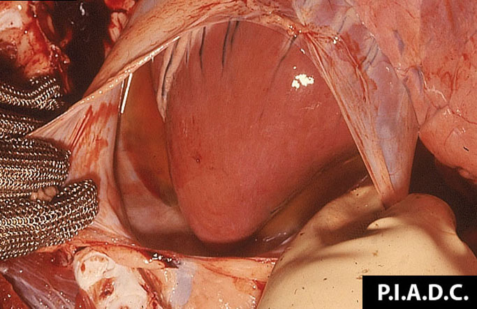
Description:
Horse, heart. The pericardial sac contains excessive, slightly turbid straw-colored fluid (hydropericardium).
Photo ID: AHS_004

Description:
Horse, heart. There are many subendocardial hemorrhages.
Photo ID: AHS_005
Description:
Horse, skeletal muscle. There is marked intermuscular edema.
Photo ID: AHS_006
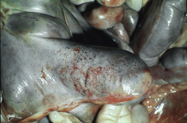
Description:
Horse, cecum. There are serosal petechiae on the apex of the cecum.
Photo ID: AHS_007