Disease Images

Disease Images: Vesicular Stomatitis

Additional resources for Vesicular Stomatitis


Description:
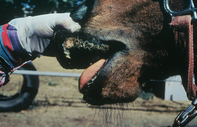
Horse, mouth. There is extensive erosion of the lip at the mucocutaneous junction.

Credit: PIADC
Photo ID: VS_001

Description:
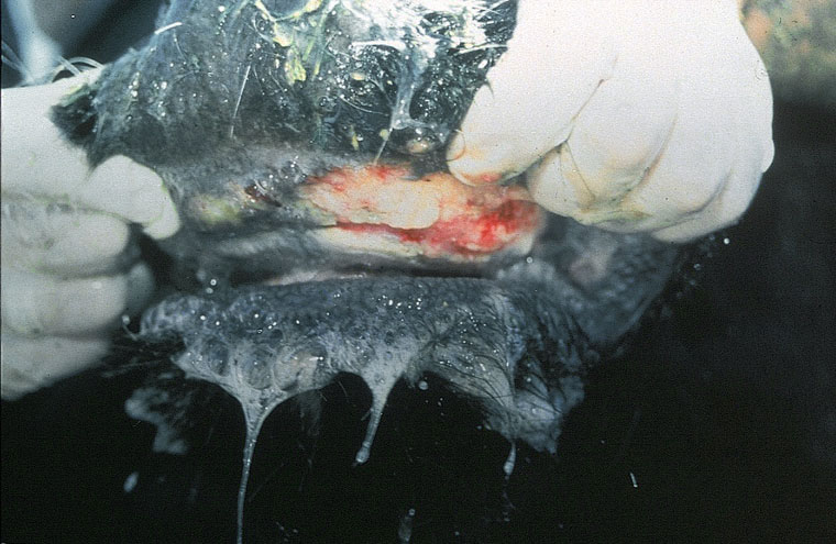
Bovine, oral. There is extensive ulceration of the dental pad, and severe salivation.

Credit: PIADC
Photo ID: VS_002

Description:
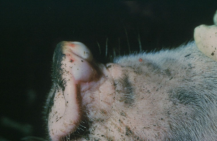
Pig, skin. There is a large vesicle (bulla) on the dorsal snout.

Credit: Iowa State University, College of Veterinary Medicine
Photo ID: VS_003

Description:
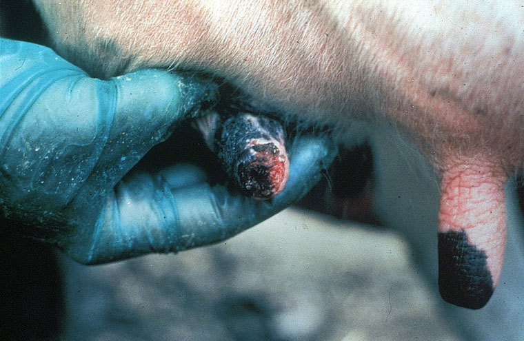
Bovine, skin. The distal teat is severely eroded and hemorrhagic.

Credit: Iowa State University, College of Veterinary Medicine
Photo ID: VS_004

To use images and associated descriptions contained on this website, please contact the CFSPH.

These images were annotated by Dr. Steve Sorden and Dr. Claire Andreasen and funding was provided by a USDA Higher Education Challenge Grant in collaboration with the Iowa State University Department of Veterinary Pathology, Center for Food Security and Public Health (CFSPH), Armed Forces Institute of Pathology (AFIP), and Plum Island Animal Disease Center (PIADC).