Disease Images
Disease Images
Disease Images: Tularemia
Additional resources for Tularemia
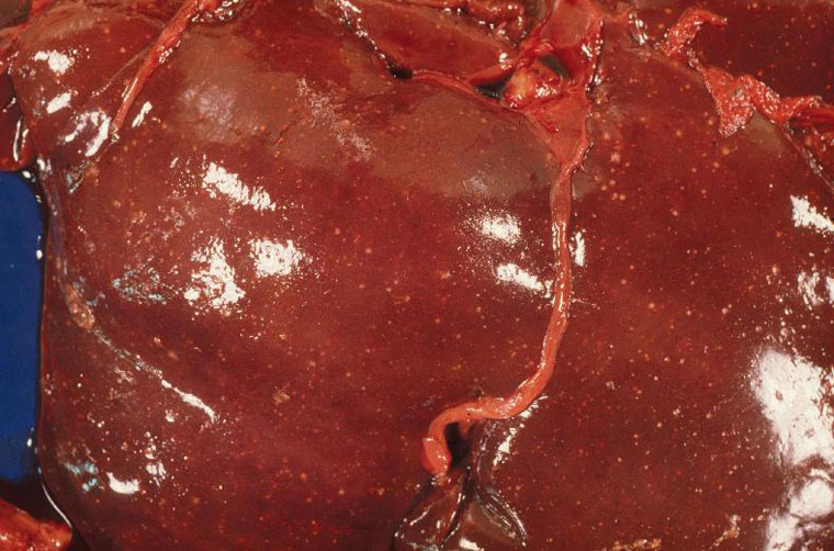
Description:
Beaver, liver. There are disseminated small pale foci of necrotizing hepatitis.
Photo ID: TUL_001
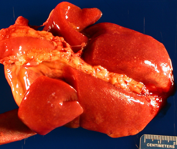
Description:
Cat, lung. Numerous <1 mm diameter pale foci are disseminated throughout all lung lobes.
Photo ID: TUL_002
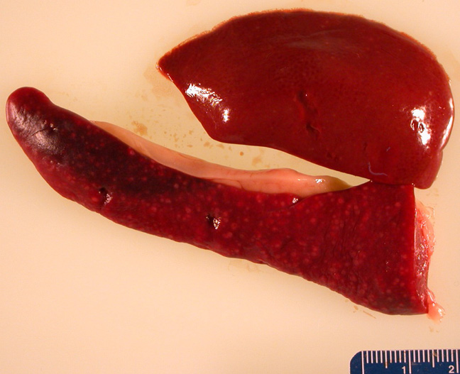
Description:
Cat, spleen and liver. Numerous ~1 mm diameter pale foci are disseminated throughout the spleen; fewer pale foci are discernable in the liver lobe.
Photo ID: TUL_003
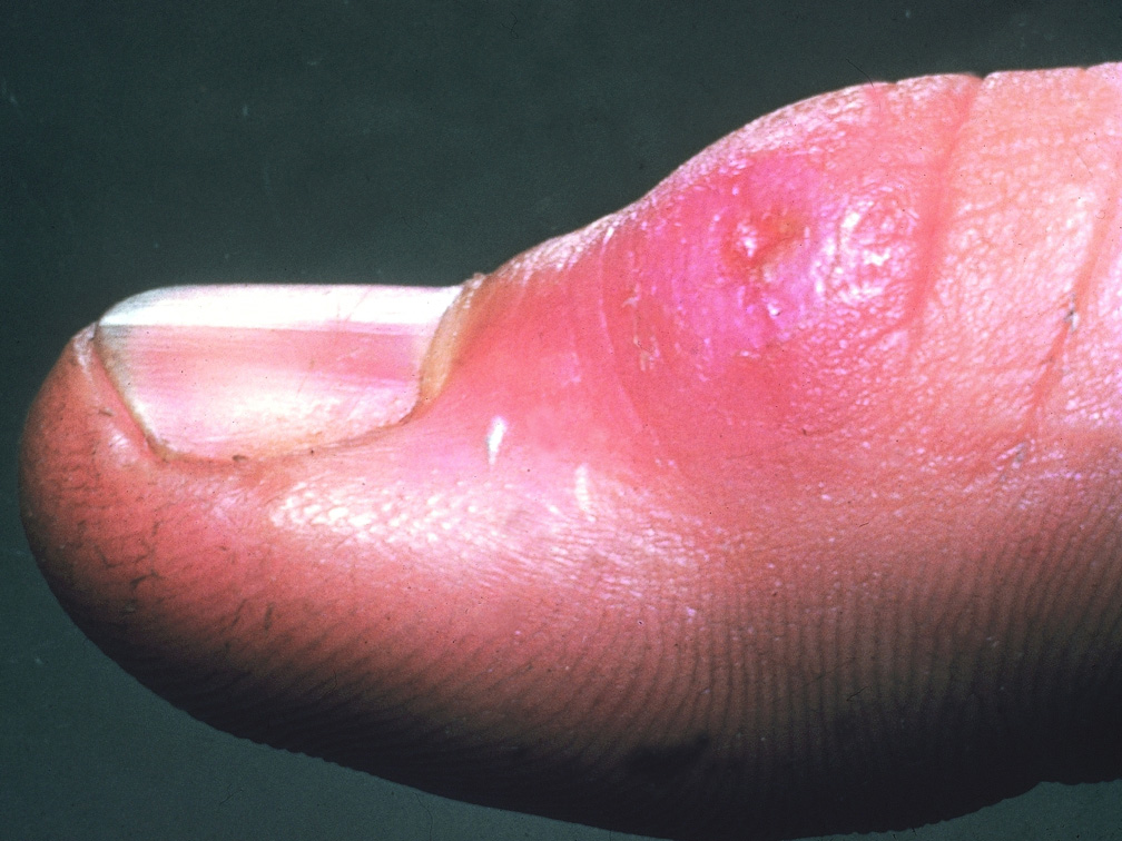
Description:
Human, skin. There is an ulcerated papule over the interphalangeal joint.
Photo ID: TUL_004