Disease Images

Disease Images: Trypanosomiasis (African)

Additional resources for Trypanosomiasis (African)


Description:
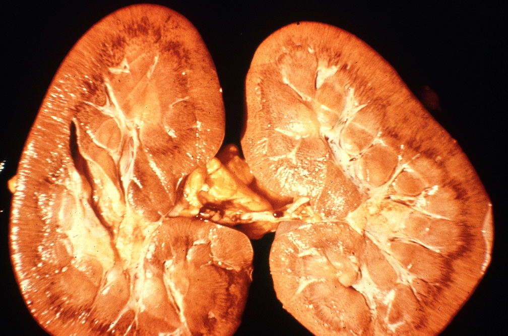
Horse, kidney. Cortex is pale and there are multiple petechial hemorrhages at the corticomedullary junction.

Credit: AFIP
Photo ID: TRY_001

Description:
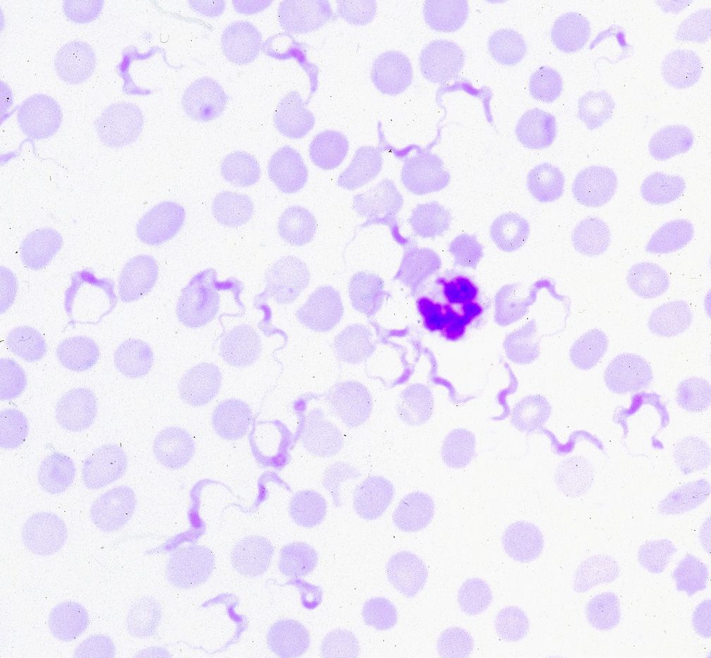
Rodent, blood smear. Numerous flagellated trypanosomes can be seen among erythrocytes and a neutrophil.

Credit: AFIP
Photo ID: TRY_002

To use images and associated descriptions contained on this website, please contact the CFSPH.

These images were annotated by Dr. Steve Sorden and Dr. Claire Andreasen and funding was provided by a USDA Higher Education Challenge Grant in collaboration with the Iowa State University Department of Veterinary Pathology, Center for Food Security and Public Health (CFSPH), Armed Forces Institute of Pathology (AFIP), and Plum Island Animal Disease Center (PIADC).