Disease Images
Disease Images
Disease Images: Theileriosis
Additional resources for Theileriosis
Description:
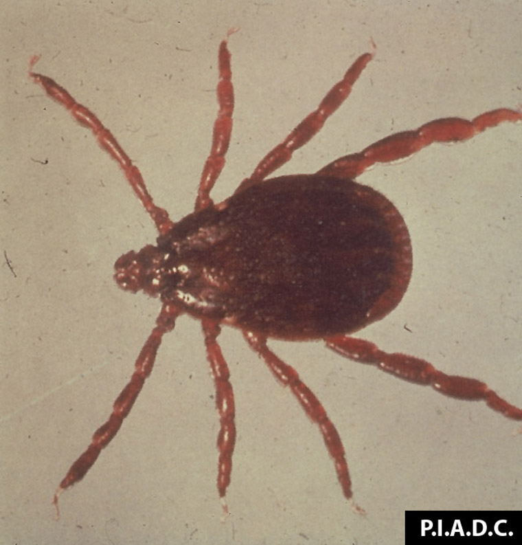
Rhipicephalus appendiculatus - brown ear tick, vector of Theileriosis.
Photo ID: THE_001
Description:
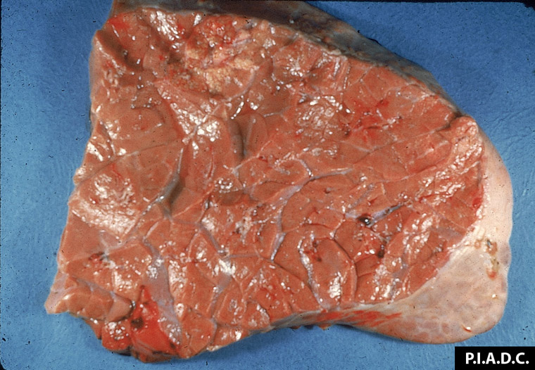
Bovine, lung. The lung tissue is diffusely tan-brown, and lobules are noncollapsed and rubbery (interstitial pneumonia).
Photo ID: THE_002

Description:
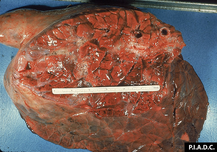
Bovine, lung. Lung tissue is noncollapsed, contains multiple foci of hemorrhage, and there is fluid/foam within interlobular septa and bronchi.
Photo ID: THE_003
Description:
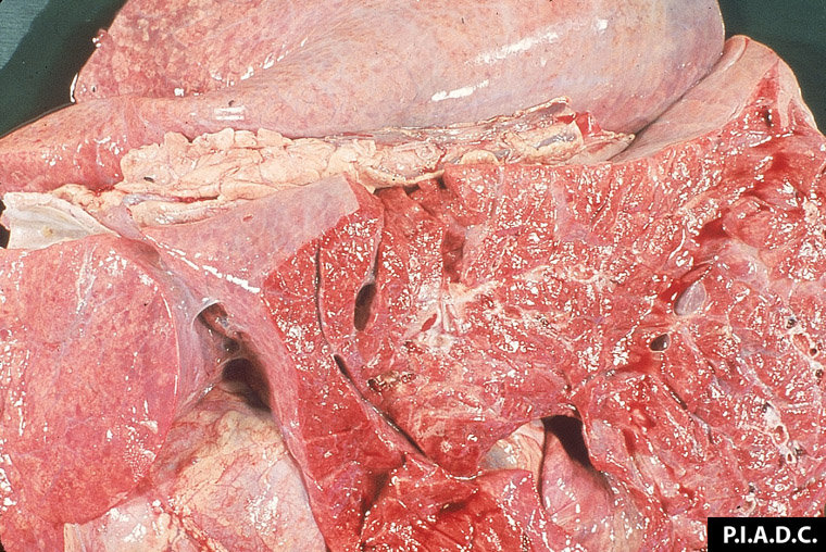
Bovine, lung. The lobules are noncollapsed (rubbery) and diffusely tan-brown, and interlobular septa are markedly expanded due to edema and emphysema.
Photo ID: THE_004
Description:
Bovine, lungs. Lungs are diffusely noncollapsed, and there is moderate interlobular edema (interstitial pneumonia).
Photo ID: THE_005
Description:
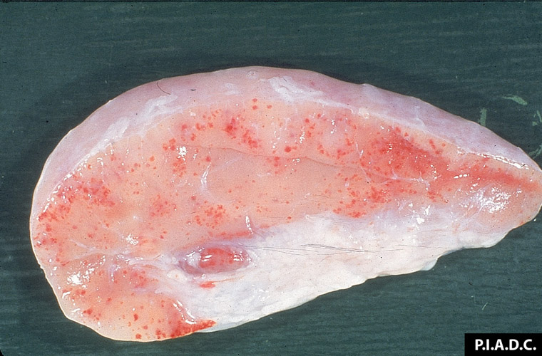
Bovine, popliteal lymph node. The node is enlarged and diffusely pale, and contains numerous petechiae.
Photo ID: THE_006
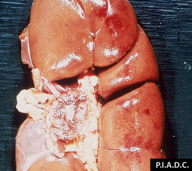
Description:
Bovine, kidney. There are multiple petechiae on the surface of the cortex. The lymph node near the hilus is markedly enlarged.
Photo ID: THE_007
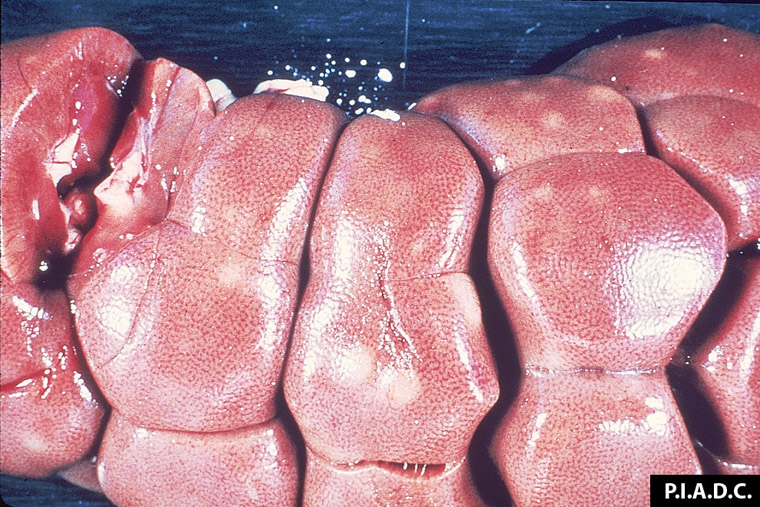
Description:
Bovine, kidney. The multiple pale foci on the cortical surface are lymphoid infiltrates.
Photo ID: THE_008
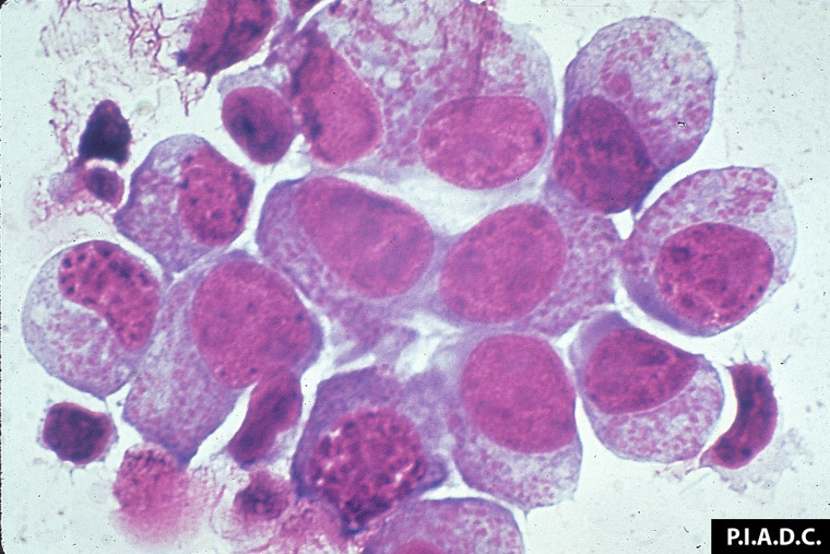
Description:
Bovine lymphoblasts contain intracytoplasmic Theileria parva.
Photo ID: THE_009