Disease Images
Disease Images
Disease Images: Sheep and Goat Pox
Additional resources for Sheep and Goat Pox
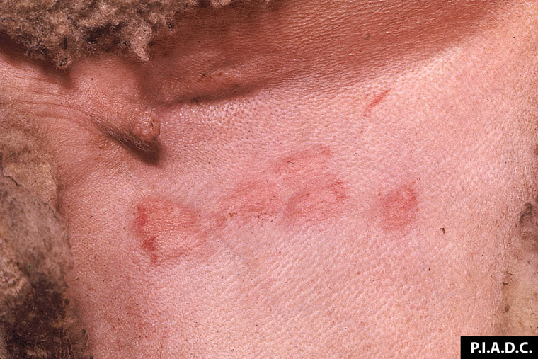
Description:
Sheep, inguinal skin. Several coalescing macules contain petechiae.
Photo ID: SGP_001
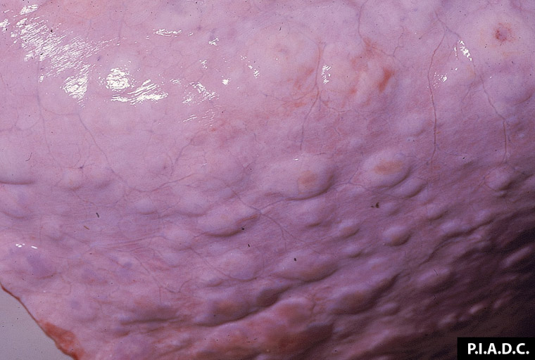
Description:
Sheep, inguinal skin. There are several coalescing macules.
Photo ID: SGP_002
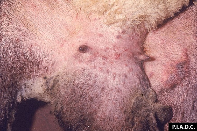
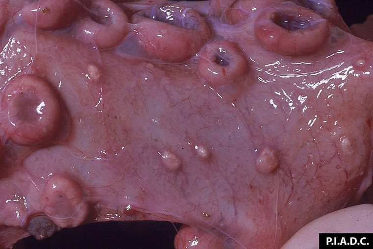
Description:
Sheep, scrotum. There are multiple papules on the scrotum and adjacent inguinal skin.
Photo ID: SGP_003
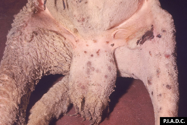
Description:
Sheep, scrotum and inguinal skin. There are multple red brown papules. There are two hemorrhagic ulcers on the medial aspect of the stifle.
Photo ID: SGP_004
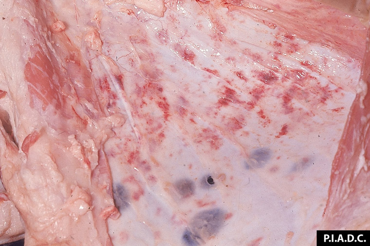
Description:
Sheep, subcutis. There are numerous hemorrhages, and several dark red round foci of hemorrhage and necrosis (beneath cutaneous pox).
Photo ID: SGP_005
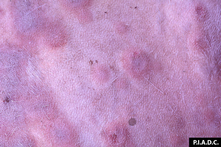
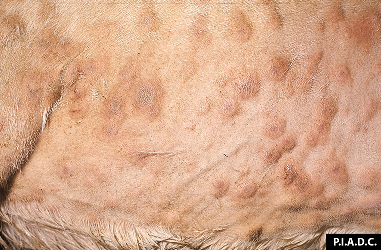
Description:
Goat, skin. Pox are coalescing red papules with central, slightly depressed, pale (necrotic) areas.
Photo ID: SGP_006
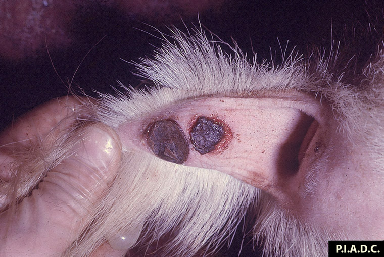
Description:
Goat. Two pox on the ventral tail have dessicated, dark red, undermined (necrotic and sloughing) centers.
Photo ID: SGP_007
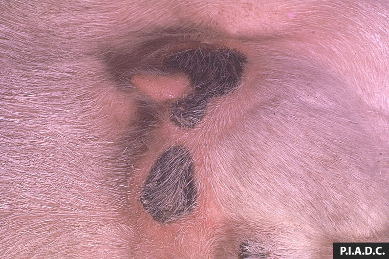
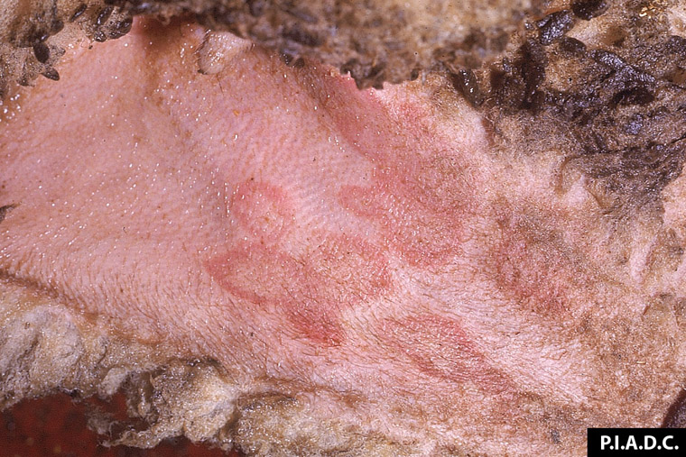
Description:
Goat, udder. The skin contains two sharply demarcated necrotic foci (subacute pox).
Photo ID: SGP_008
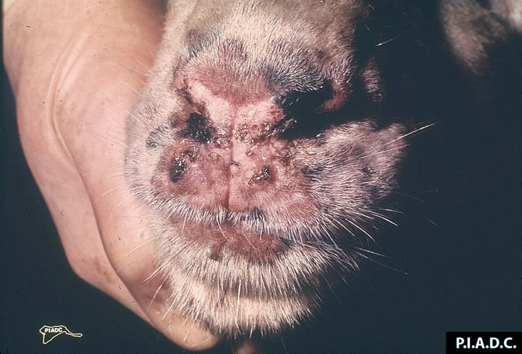
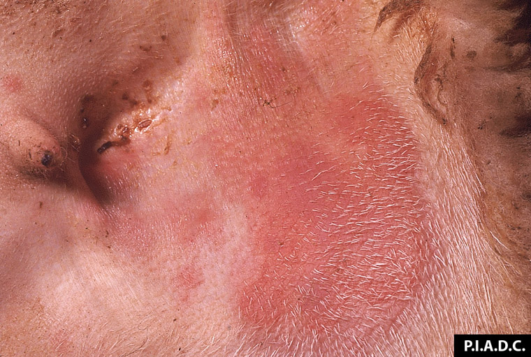
Description:
Goat, muzzle. The muzzle contains several papules and is partially covered by hemorrhagic nasal exudate.
Photo ID: SGP_009
Description:
Sheep, skin. Several coalescing pox have pale tan (necrotic) centers.
Photo ID: SGP_010
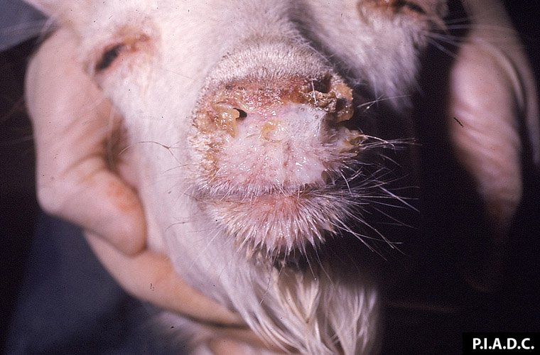
Description:
Goat. Abundant thick nasal exudate covers the muzzle and partially occludes the nares.
Photo ID: SGP_011
Description:
Goat, skin. There are multiple coalescing papules (pox) that often have tan, dry (necrotic) centers.
Photo ID: SGP_012
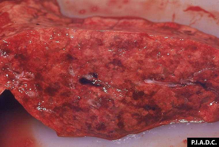
Description:
Small ruminant, lung. There are numerous, small, coalescing, red-tan, consolidated foci (pneumonia).
Photo ID: SGP_013
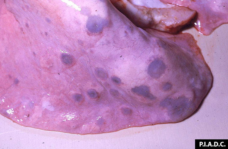
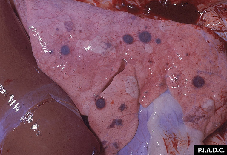
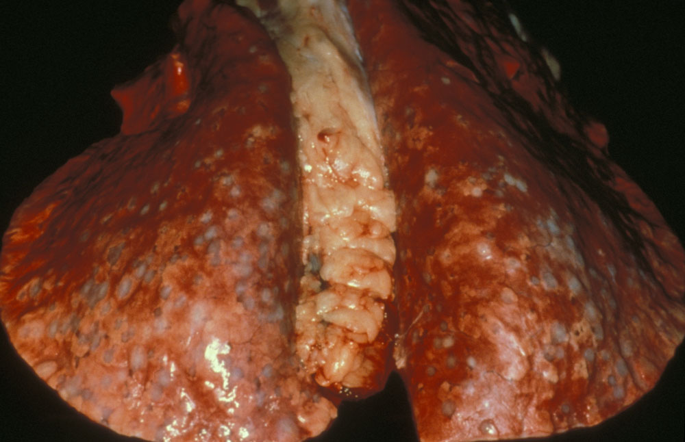
Description:
Small ruminant, lungs. The lungs contain multiple discrete tan to red-brown nodules (multifocal interstitial pneumonia). Mediastinal lymph nodes are enlarged.
Photo ID: SGP_014
Description:
Small ruminant, lung. There are multiple red-brown consolidated foci (multifocal pneumonia).
Photo ID: SGP_015
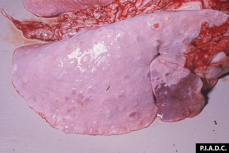
Description:
Small ruminant, lung. Numerous, slightly raised, pale tan to red-brown nodules (foci of consolidation) are scattered throughout the lung; the locally extensive cranioventral red-brown consolidation is likely secondary bacterial bronchopneumonia.
Photo ID: SGP_016
Description:
Small ruminant, lung. There are numerous raised pale nodules (multifocal pneumonia).
Photo ID: SGP_017
Description:
Small ruminant, lung. There are multiple discrete, round, red-brown foci of consolidation (pneumonia).
Photo ID: SGP_018
Description:
Sheep, lung. The numerous widely disseminated discrete round tan foci are foci of pneumonia; a few have pale (necrotic) centers.
Photo ID: SGP_019
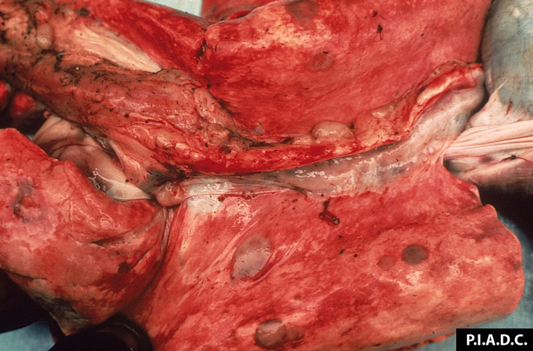
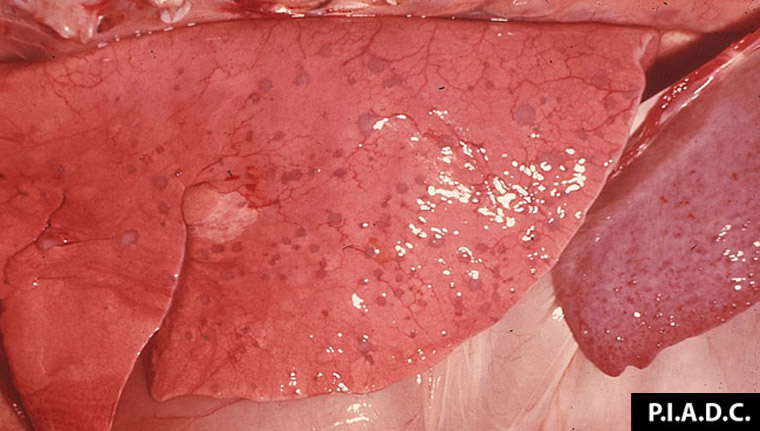
Description:
Goat, lung. There are multiple coalescing tan foci of consolidation (pneumonia), and the adjacent lymph node is markedly enlarged.
Photo ID: SGP_020
Description:
Small ruminant, uterus. The endometrium contains several tan papules (pox) among the caruncles.
Photo ID: SGP_021
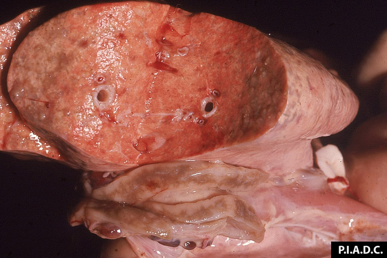
Description:
Sheep, lungs. Lungs with diffuse granulomatous nodules.
Photo ID: SGP_022