Disease Images

Disease Images: Sarcocystosis

Additional resources for Sarcocystosis


Description:
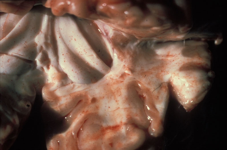
Bovine, brain. Brain with multifocal diffuse petecchia that are more prominent in the white matter.

Credit: AFIP
Photo ID: SAR_001

Description:
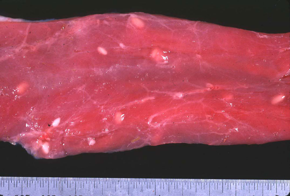
Sheep, esophagus. Esophogeal muscle with white oval multifocal cysts (sarcocytes) of sarcocystosis.

Credit: Dr. C. Leathers, Washington State University, Noah’s Arkive
Photo ID: SAR_002

To use images and associated descriptions contained on this website, please contact the CFSPH.

These images were annotated by Dr. Steve Sorden and Dr. Claire Andreasen and funding was provided by a USDA Higher Education Challenge Grant in collaboration with the Iowa State University Department of Veterinary Pathology, Center for Food Security and Public Health (CFSPH), Armed Forces Institute of Pathology (AFIP), and Plum Island Animal Disease Center (PIADC).