Disease Images
Disease Images
Disease Images: Lumpy Skin Disease
Additional resources for Lumpy Skin Disease
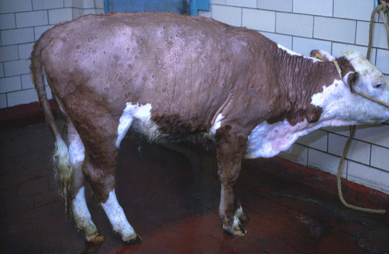
Description:
Bovine, skin. There are disseminated cutaneous papules.
Photo ID: LSD_001
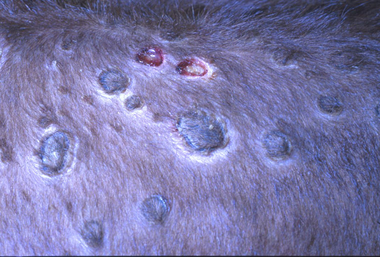
Description:
Bovine, skin. There are disseminated cutaneous papules with necrotic centers (sitfasts).
Photo ID: LSD_002
Description:
Bovine, skin. Necrotic centers (sitfasts) of two of these papules have sloughed.
Photo ID: LSD_003
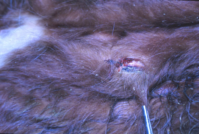
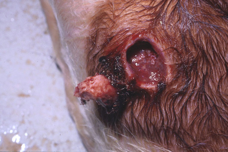
Description:
Bovine, skin. Removal of the necrotic center (sitfast) of a papule.
Photo ID: LSD_004
Description:
Bovine, skin. There is hemorrhagic exudate subjacent to the necrotic center (sitfast) of a papule.
Photo ID: LSD_005
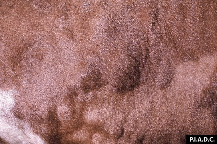
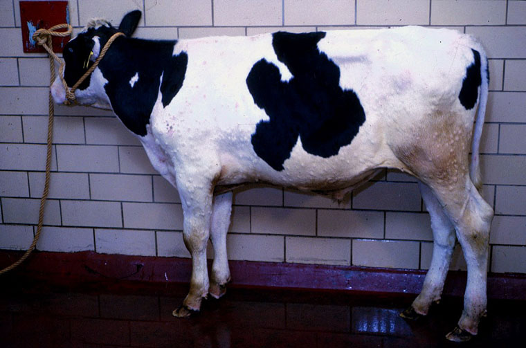
Description:
Bovine, skin. Multiple subcutaneous nodules elevate the skin.
Photo ID: LSD_006
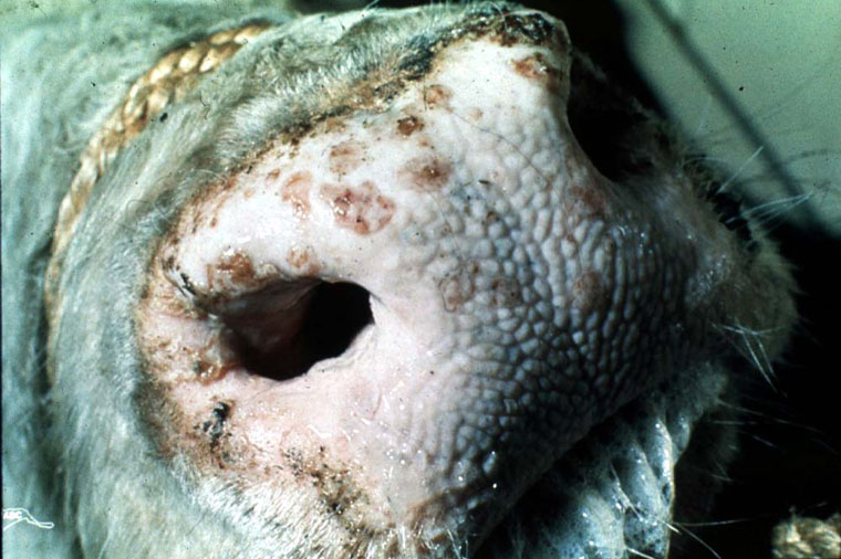
Description:
Bovine, muzzle. There are multiple sharply-demarcated slightly raised papules, often with eroded surfaces, that extend into the nares.
Photo ID: LSD_007
Description:
Bovine, nasal turbinate. Early pox lesions are slightly pale round foci rimmed by petechiae.
Photo ID: LSD_008
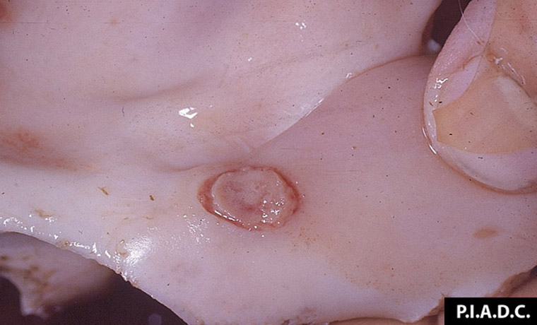
Description:
Bovine, nasal turbinate. The centers of well-developed pox are necrotic.
Photo ID: LSD_009
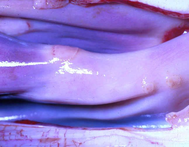
Description:
Bovine, nasal turbinate. Nasal mucosa contains several macules with hyperemic margins.
Photo ID: LSD_010
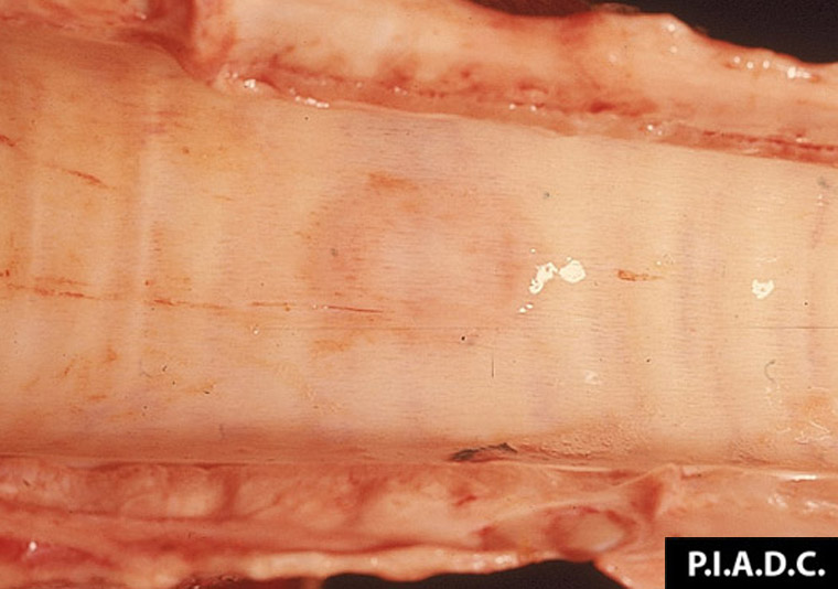
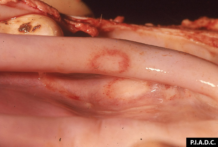
Description:
Bovine, trachea. The mucosa contains a poorly demarcated round focus rimmed by mild hemorrhage (early pox lesion).
Photo ID: LSD_011
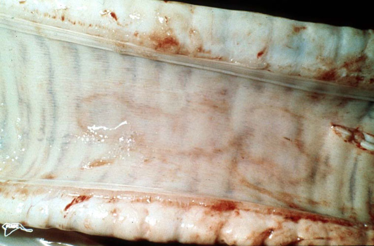
Description:
Bovine, trachea. Two coalescing mucosal macules have hyperemic margins.
Photo ID: LSD_012
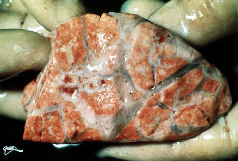
Description:
Bovine, lung. There is marked generalized interlobular edema, and there is a small cluster of red nodules on the left side of the specimen.
Photo ID: LSD_013