Disease Images
Disease Images
Disease Images: Ehrlichiosis
Additional resources for Ehrlichiosis
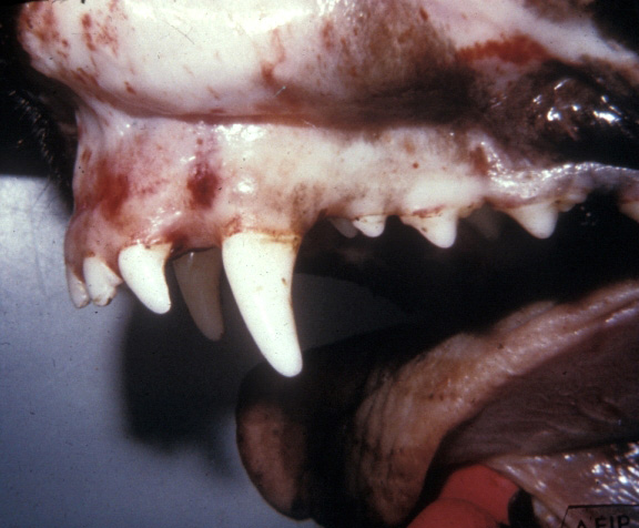
Description:
Dog, oral mucosa, Ehrlichia canis. There are multiple petechiae and ecchymoses on the gingival and buccal mucosa.
Photo ID: EHR_001
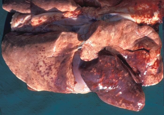
Description:
Dog, lungs, Ehrlichia canis. There are multiple coalescing hemorrhages on the pleural surface; the right middle lobe is also edematous.
Photo ID: EHR_002
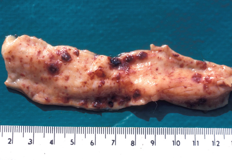
Description:
Dog, small intestine, Ehrlichia canis. There are multiple mucosal petechiae that coalesce to form larger, raised foci of hemorrhage.
Photo ID: EHR_003