Disease Images
Disease Images
Disease Images: Classical Swine Fever
Additional resources for Classical Swine Fever
Description:
Pig, kidney. The cortex contains multiple petechiae and pale infarcts surrounded by hemorrhage.
Photo ID: CSF_001
Description:
Pig, kidney. The cortex contains multiple petechiae and pale infarcts surrounded by hemorrhage.
Photo ID: CSF_002
Description:
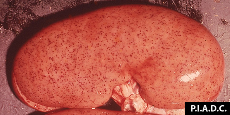
Pig, kidney. There are numerous disseminated cortical petechiae ("turkey egg kidney").
Photo ID: CSF_003
Description:
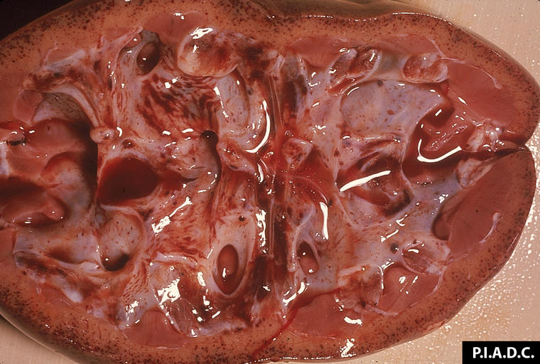
Pig, kidney. The cortex contains disseminated petechiae. Calyces are moderately dilated (hydronephrosis) and also contain hemorrhages.
Photo ID: CSF_004
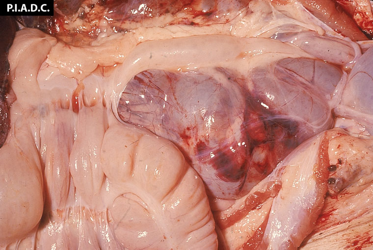
Description:
Pig, retropharyngeal lymph node. The lymph node is markedly enlarged and hemorrhagic; the tonsil contains multiple poorly demarcated hemorrhages.
Photo ID: CSF_005
Description:
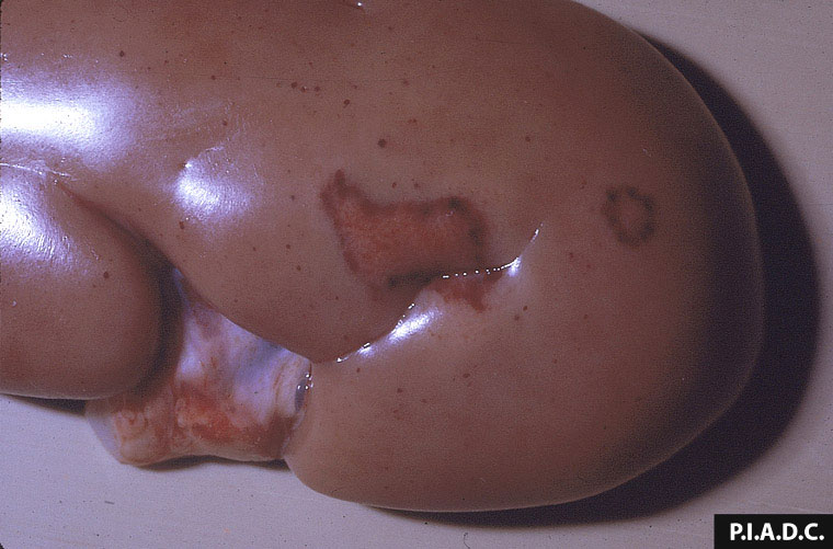
Pig, kidney. There is extensive hemorrhage on the cortical surface.
Photo ID: CSF_006
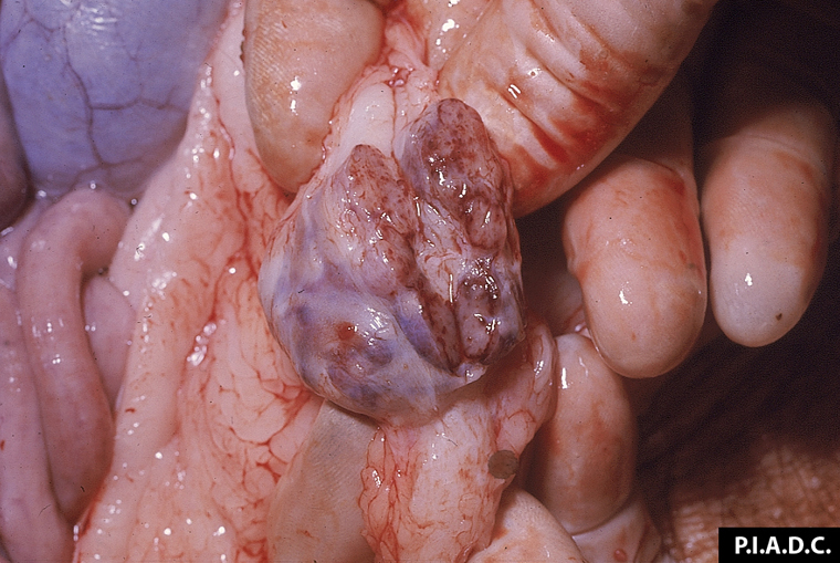
Description:
Pig, inguinal lymph node. There are petechial and peripheral (medullary sinus) hemorrhages.
Photo ID: CSF_007
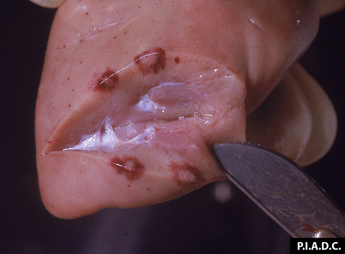
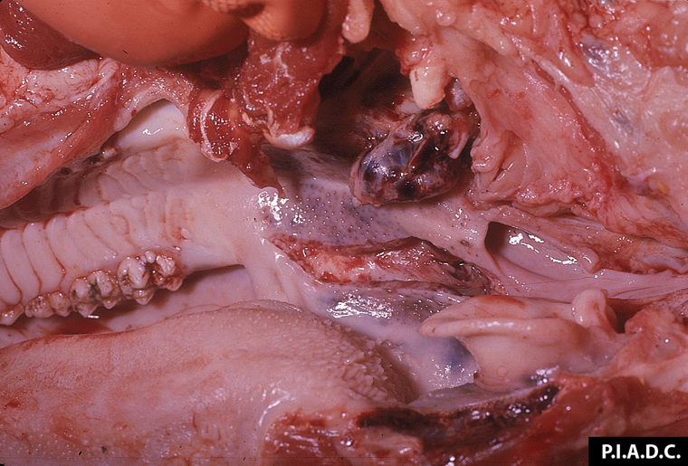
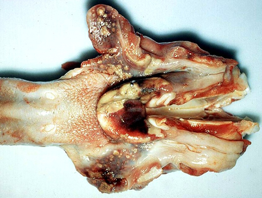
Description:
Pig, pharynx and larynx. There are coalescing foci of petechial hemorrhage (and necrosis) in the palatine tonsils and adjacent pharyngeal and laryngeal mucosa.
Photo ID: CSF_008

Description:
Pig, lungs. There are numerous disseminated pleural petechiae, and there is mild interlobular edema.
Photo ID: CSF_009
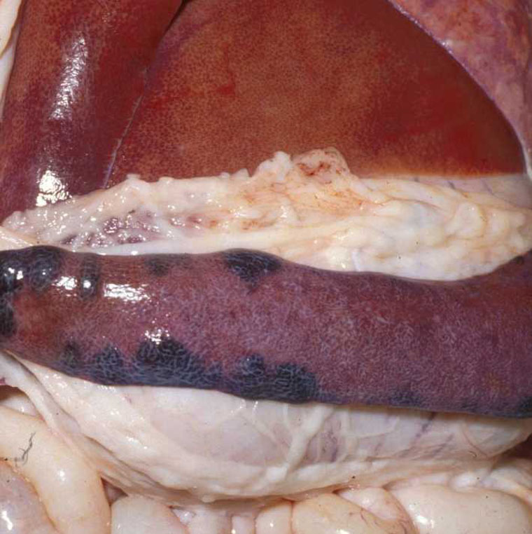
Description:
Pig, spleen. There are multiple coalescing, swollen, dark red infarcts along the margins.
Photo ID: CSF_010
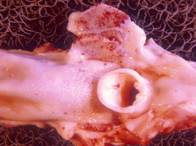
Description:
Pig, tonsil. The epiglottis and the bisected palatine tonsil contain multiple tan foci of necrosis.
Photo ID: CSF_011
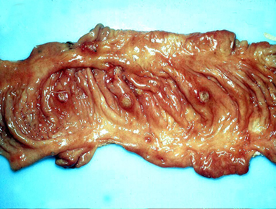
Description:
Porcine, colon. The mucosa is reddened and contains multiple discrete ("button") ulcers surrounded by zones of hemorrhage.
Photo ID: CSF_012