Disease Images
Disease Images
Disease Images: Campylobacteriosis
Additional resources for Campylobacteriosis
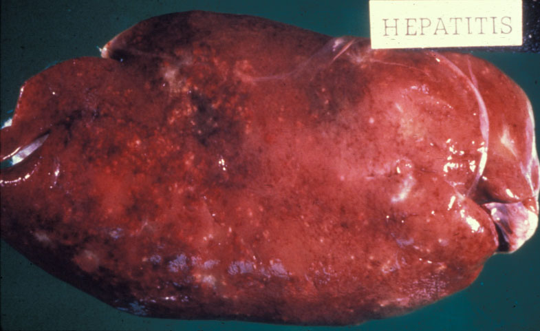
Description:
Avian, liver. Swollen liver with rounded edges and multifocal white lesions due to Campylobacterosis.
Photo ID: CAM_001
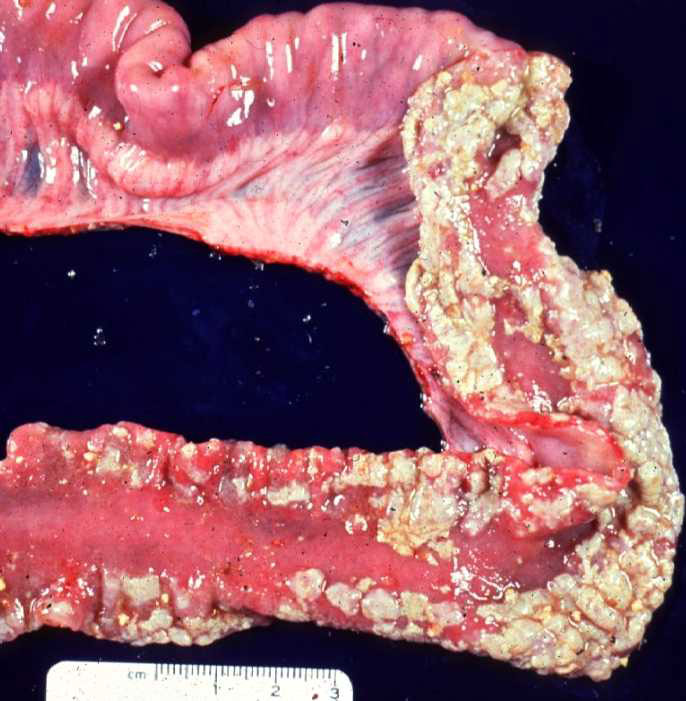
Description:
Pig, small intestine. White to tan multifocal luminal exudate within the small intestine.
Photo ID: CAM_002
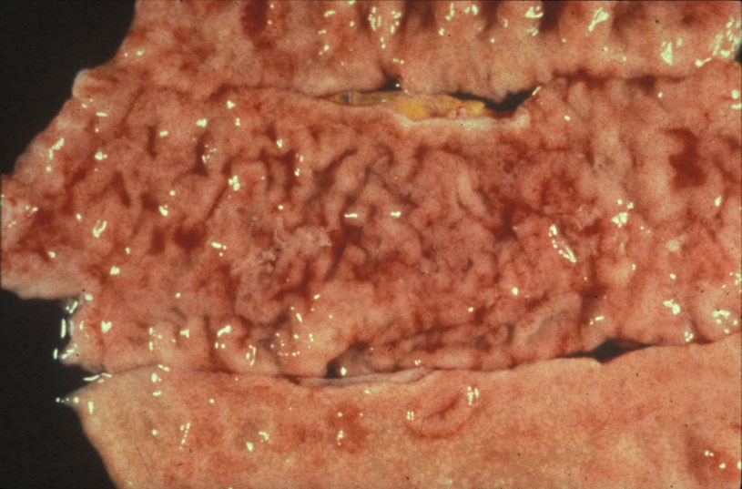
Description:
Primate, colon. Mucosal edema, muco-hemorrhagic exudate, and thickened folds of the colon.
Photo ID: CAM_003
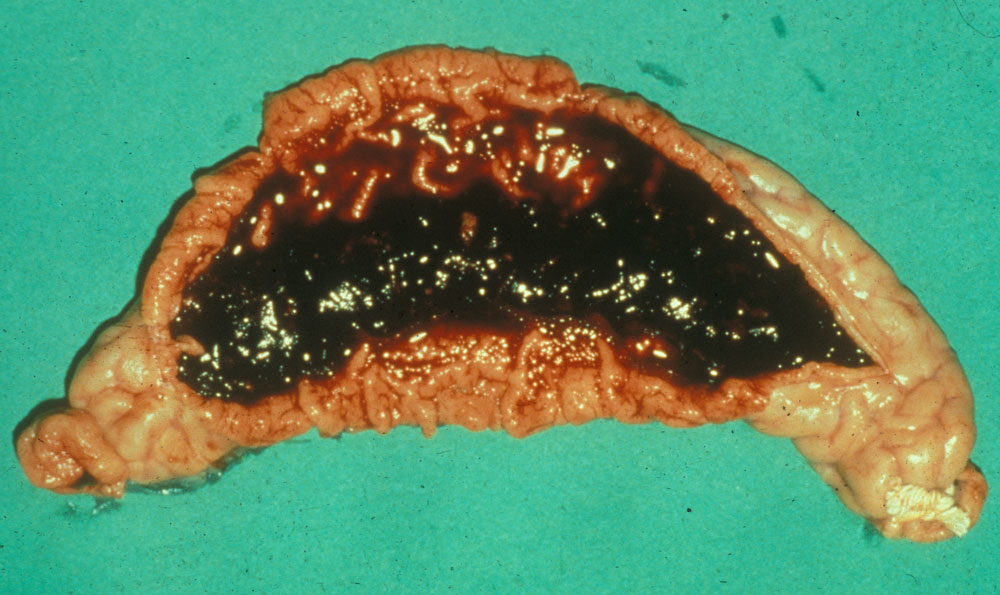
Description:
Pig, small intestine. Hemorrhage in the lumen of the small intestine with thickened mucosal folds.
Photo ID: CAM_004