Disease Images
Disease Images
Disease Images: Bluetongue
Additional resources for Bluetongue
Description:
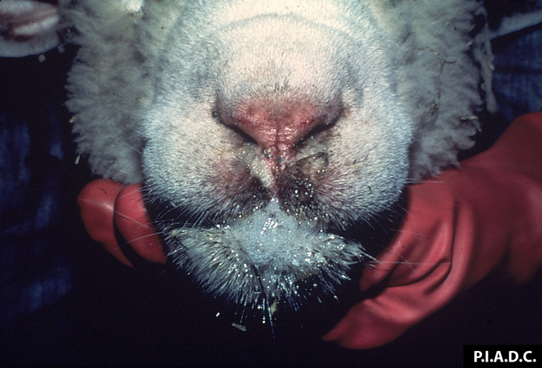
Sheep. There is bilateral nasal exudate, erosion of the nasal planum, and excessive salivation.
Photo ID: BT_001
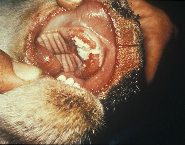
Description:
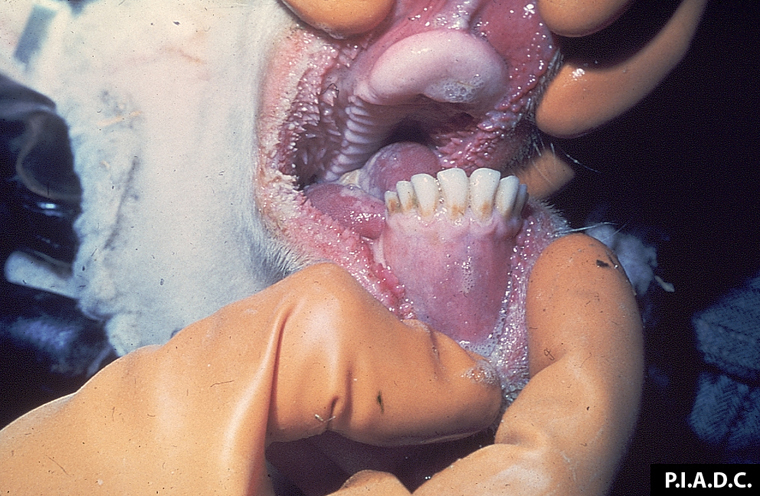
Sheep, mouth. There is linear erosion and reddening of the right buccal mucosa.
Photo ID: BT_002
Description:
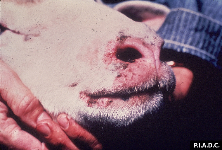
Sheep. There are multiple erosions and crusts on the muzzle and lips.
Photo ID: BT_003
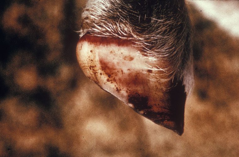
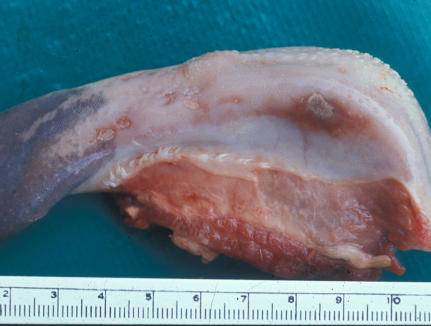
Description:
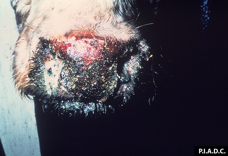
Bovine. The muzzle is covered by an adherent crust, and the underlying (eroded) tissue is hyperemic.
Photo ID: BT_004
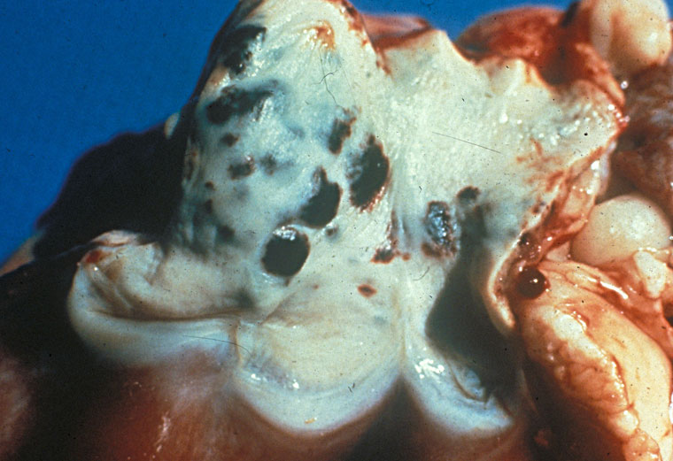
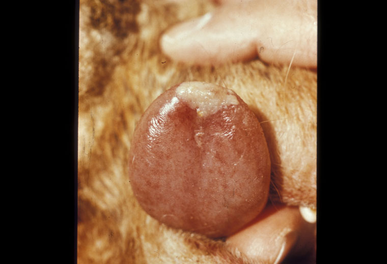
Description:
Sheep, mouth. Most of the dental pad is eroded; the remaining pale mucosa is necrotic.
Photo ID: BT_005
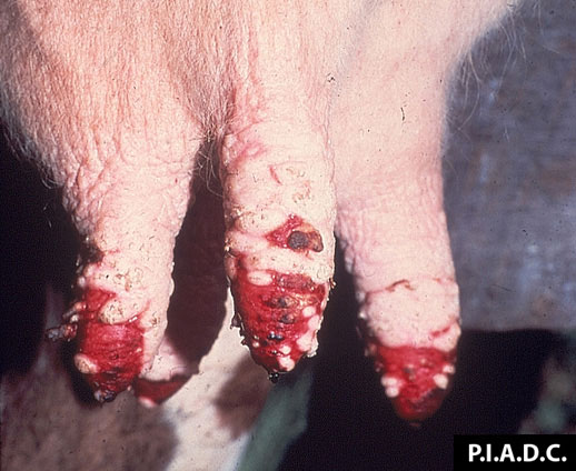
Description:
Bovine, mammary gland. There is extensive coalescing ulceration of the teat skin.
Photo ID: BT_006
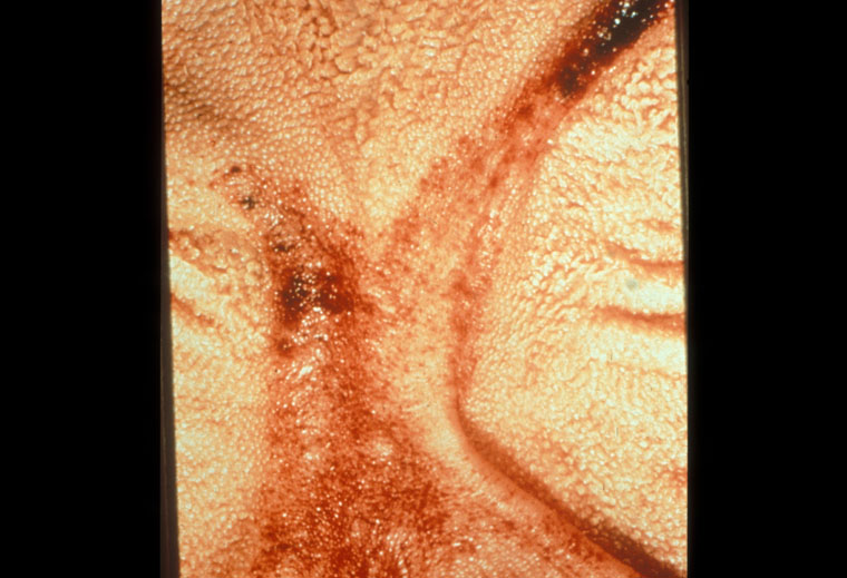
Description:
Sheep, pulmonary artery. There are multiple ecchymoses on the intimal surface.
Photo ID: BT_007
Description:
Sheep, foot. There are multiple petechiae in the hoof wall, and there is marked hyperemia of the coronary band.
Photo ID: BT_008
Description:
Sheep, tongue. The lateral mucosa contains several ulcers that are covered by exudate and surrounded by zones of hyperemia.
Photo ID: BT_009
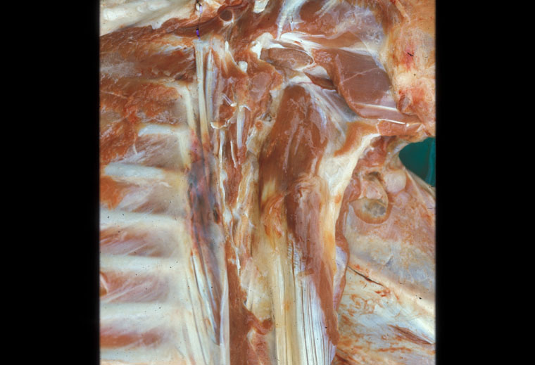
Description:
Sheep, skeletal muscle. There is a focus of hemorrhage on the left; pale areas are consistent with myodegeneration.
Photo ID: BT_010
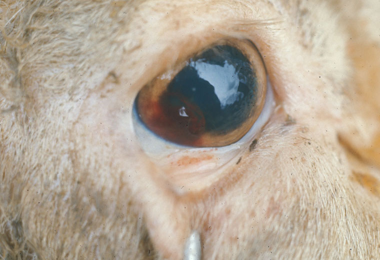
Description:
Sheep, eye. There are foci of bulbar and palpebral conjunctival hemorrhage.
Photo ID: BT_011
Description:
Sheep, tongue. There are disseminated mucosal petechiae, and a single large vesicle on the tip.
Photo ID: BT_012
Description:
Sheep, rumen. There are multiple mucosal hemorrhages centered on the pillars.
Photo ID: BT_013
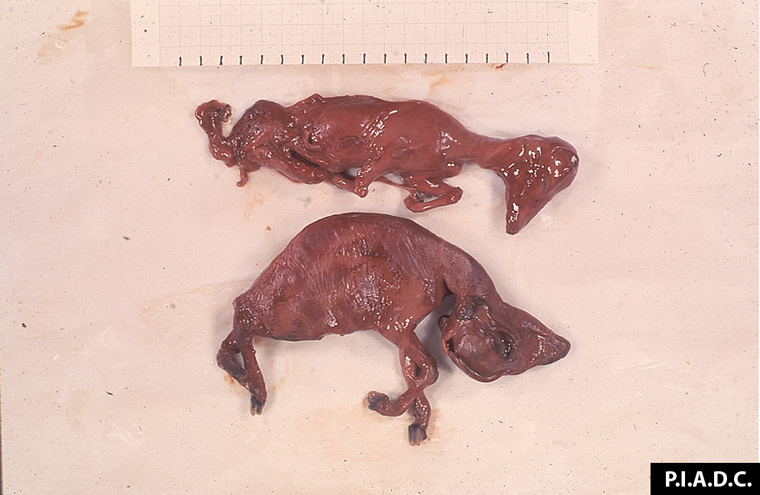
Description:
Sheep, fetuses. The larger of these aborted macerated fetuses exhibits torticollis.
Photo ID: BT_014