Disease Images

Disease Images: Rocky Mountain Spotted Fever

Additional resources for Rocky Mountain Spotted Fever
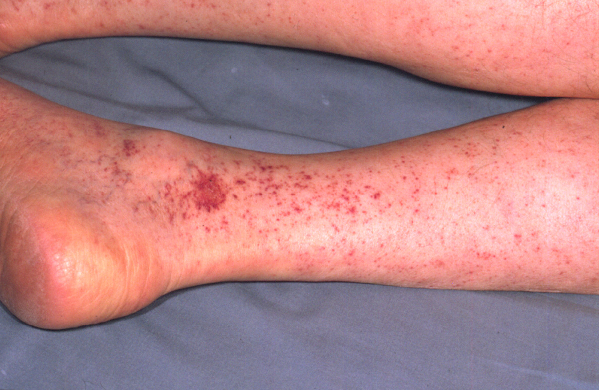

Description:
Human, legs. Disseminated cutaneous petechiae coalesce in two foci to form ecchymoses.

Credit: AFIP, National Museum of Health and Medicine
Photo ID: RMSF_001

Description:
Human, skin. There are numerous, often coalescing petechiae. A single ecchymosis exhibits desquamation and contains a dark red focus (necrosis).

Credit: AFIP, National Museum of Health and Medicine
Photo ID: RMSF_002

To use images and associated descriptions contained on this website, please contact the CFSPH.

These images were annotated by Dr. Steve Sorden and Dr. Claire Andreasen and funding was provided by a USDA Higher Education Challenge Grant in collaboration with the Iowa State University Department of Veterinary Pathology, Center for Food Security and Public Health (CFSPH), Armed Forces Institute of Pathology (AFIP), and Plum Island Animal Disease Center (PIADC).