Disease Images
Disease Images
Disease Images: Rinderpest
Additional resources for Rinderpest
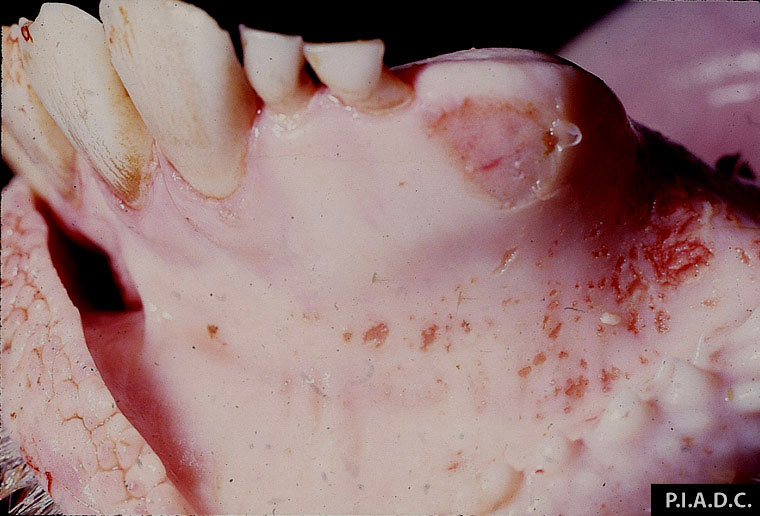
Description:
Bovine, oral mucosa. There are numerous small gingival erosions.
Photo ID: RP_001
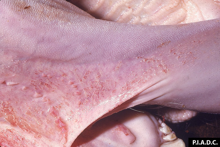
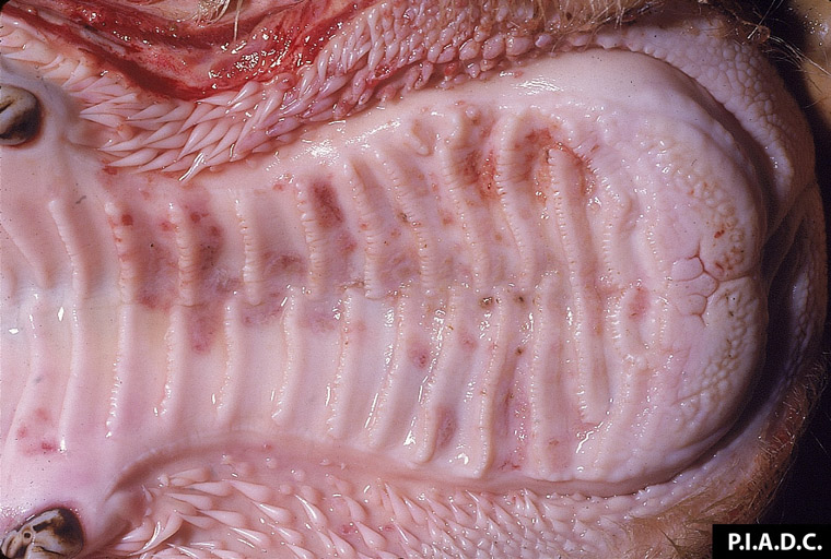
Description:
Bovine, oral mucosa. There are numerous coalescing erosions on the ventrolateral lingual mucosa.
Photo ID: RP_002
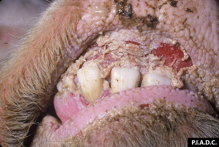
Description:
Bovine, oral mucosa. There is severe diffuse necrosis/coalescing ulceration of the dental pad; mandibular mucosa contains smaller erosions.
Photo ID: RP_003

Description:
Bovine, gingiva. There are a few small erosions.
Photo ID: RP_004
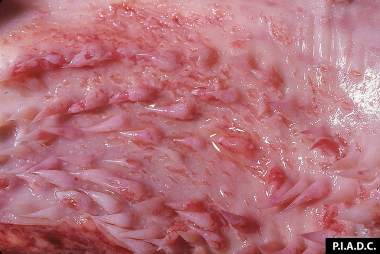
Description:
Bovine, oral mucosa. There are numerous erosions on and between the buccal papillae.
Photo ID: RP_005
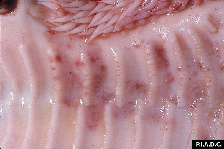
Description:
Bovine, hard palate. The mucosa contains many small, coalescing, pale to dark red erosions or foci of necrosis.
Photo ID: RP_006
Description:
Bovine, hard palate. Palate erosion.
Photo ID: RP_007

Description:
Bovine, trachea. The mucosa is hyperemic and covered by abundant mucopurulent exudate.
Photo ID: RP_008
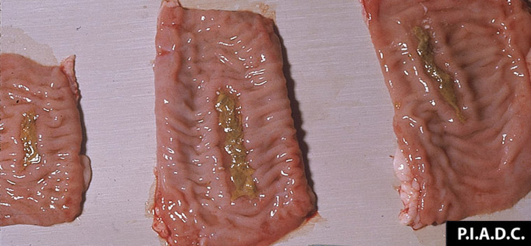
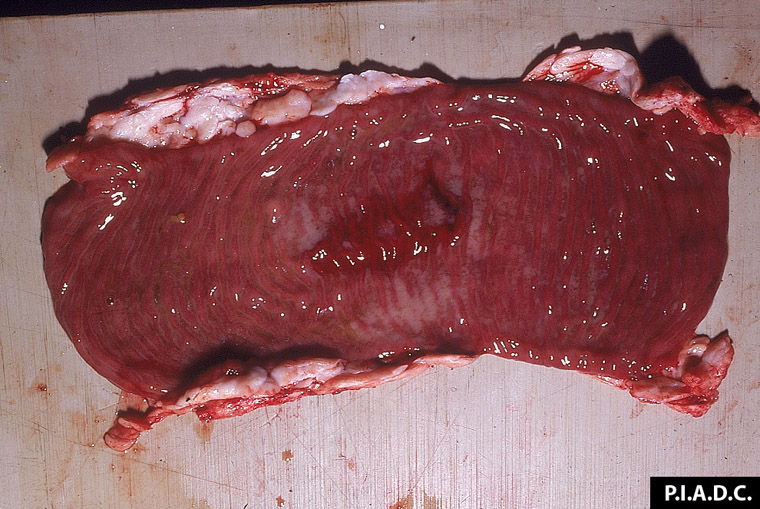
Description:
Bovine, ileum. Peyer's patches are depressed and covered by fibrinonecrotic exudate.
Photo ID: RP_009
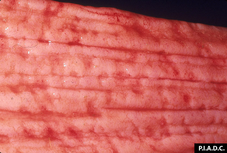
Description:
Bovine, colon. There are many petechiae on the crests of the mucosal folds, and there are several small blood clots on the mucosal surface.
Photo ID: RP_010
Description:
Bovine, ileum. The mucosa is hemorrhagic and edematous, and the Peyer's patch is depressed (necrosis).
Photo ID: RP_011
Description:
Bovine, colon. The mucosa is edematous and contains many small hemorrhages and shallow erosions.
Photo ID: RP_012
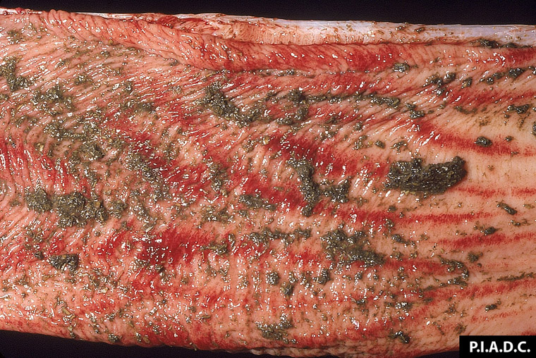
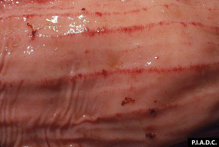
Description:
Bovine, colon. The mucosa contains multiple longitudinal linear hemorrhages.
Photo ID: RP_013
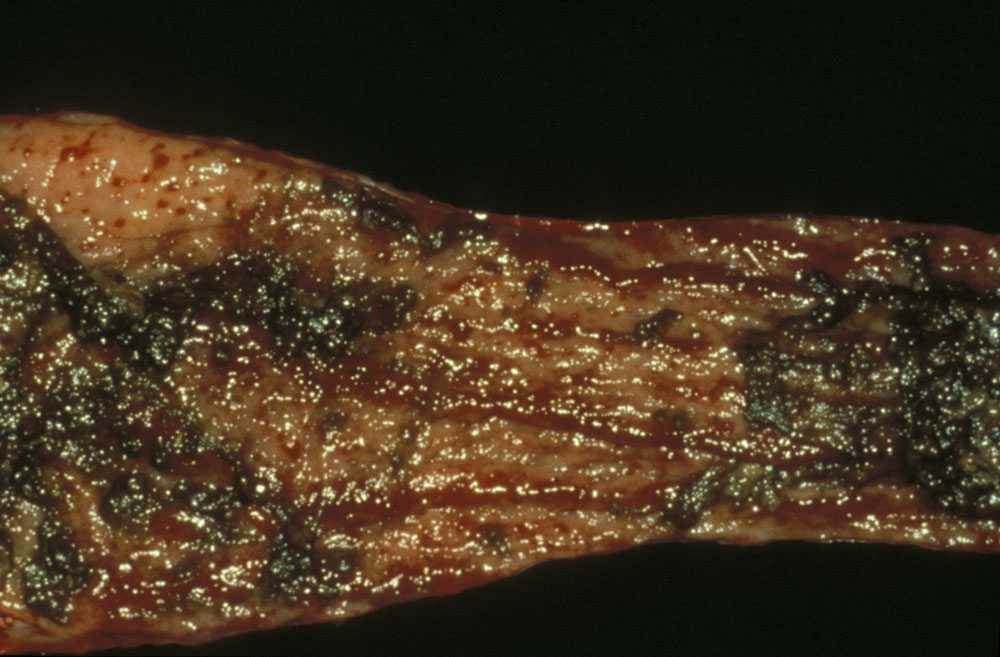
Description:
Bovine, intestine. Erosions with ulceration; dark areas of mucosal necrosis and hemorrhage.
Photo ID: RP_014