Disease Images
Disease Images
Disease Images: Newcastle Disease
Additional resources for Newcastle Disease
Description:
Chicken. The comb is markedly edematous and contains multiple foci of hemorrhage.
Photo ID: ND_001

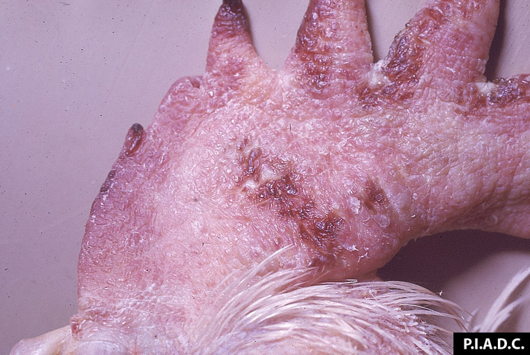
Description:
Avian, skin. There is a marked hemorrhage of the comb, wattle, and adjacent skin.
Photo ID: ND_002
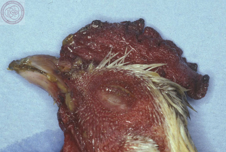
Description:
Avian, skin. There is marked hemorrhage of the comb and head, with cyanosis of the margin of the comb.
Photo ID: ND_003
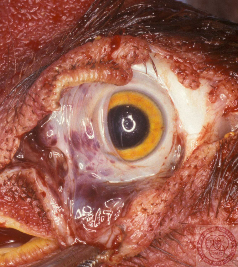
Description:
Avian, eye. Conjunctival hemorrhage is most severe in the nictitans.
Photo ID: ND_004
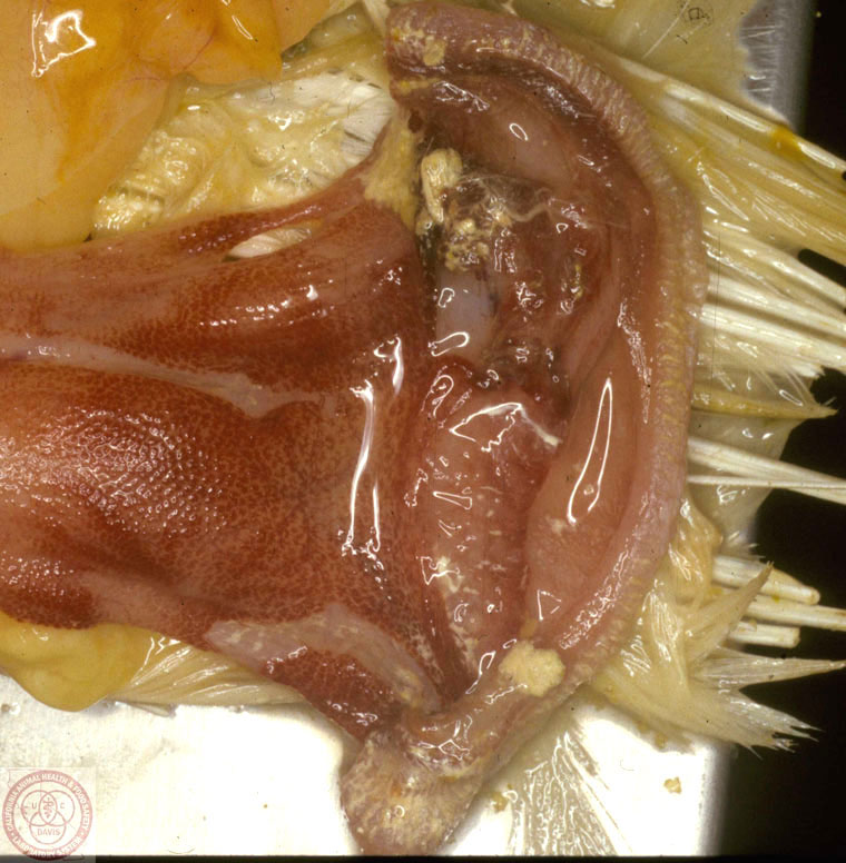
Description:
Avian, oral cavity. Numerous clumps of fibrinonecrotic exudate adhere to foci of necrosis in the oral, pharyngeal, and esophageal mucosa.
Photo ID: ND_005
Description:
Avian, trachea. Tracheal and laryngeal mucosa contain many foci of hemorrhage and small clumps of fibrinonecrotic exudate.
Photo ID: ND_006

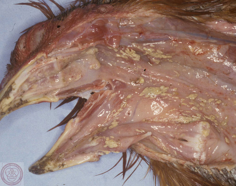
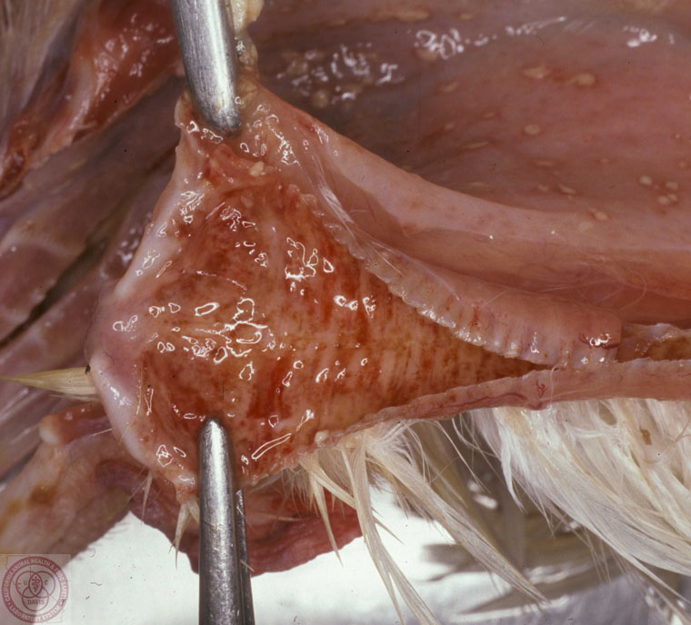
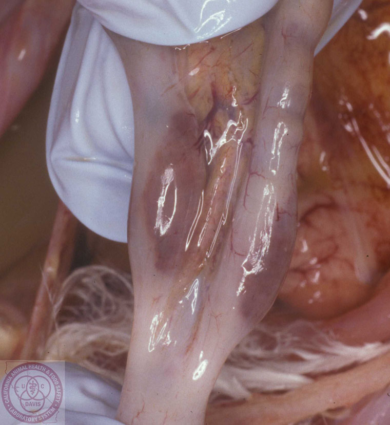
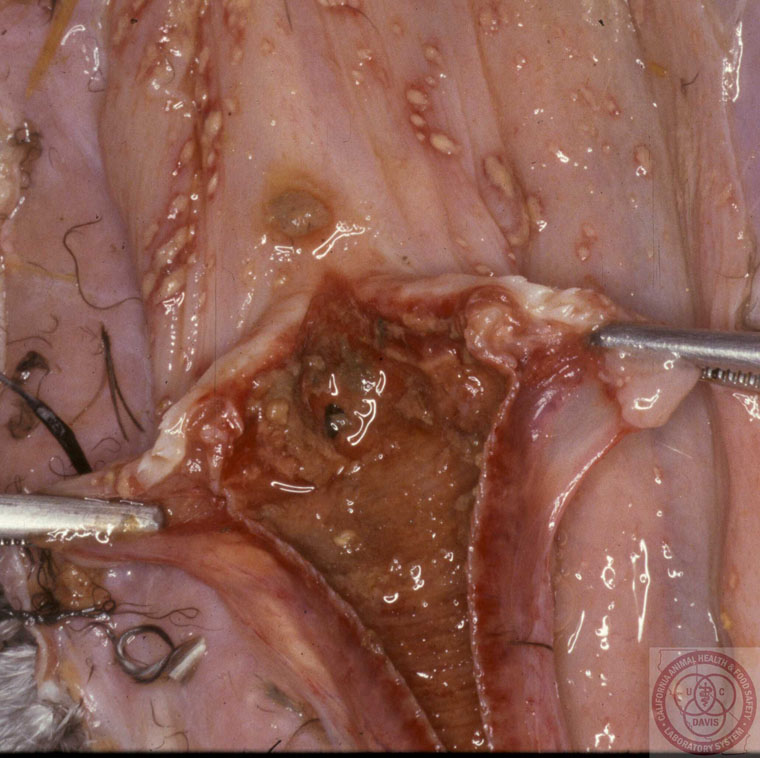
Description:
Avian, skin. There is marked subcutaneous edema in the neck, extending to the thoracic inlet.
Photo ID: ND_007
Description:
Avian, ceca. Hyperemic, necrotic cecal tonsils are visible from the serosal surface.
Photo ID: ND_008
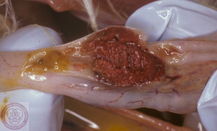
Description:
Avian, ceca. The cecal tonsil is red-brown, thickened, and friable (necrotic).
Photo ID: ND_009
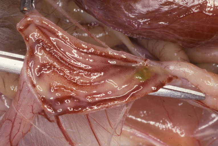
Description:
Avian, rectum. There are multiple linear mucosal hemorrhages.
Photo ID: ND_010
Description:
Avian, cloaca. The mucosa is hyperemic and contains foci of hemorrhage.
Photo ID: ND_011
Description:
Avian, colon. The mucosa contains multiple sharply demarcated foci of hemorrhage and necrosis.
Photo ID: ND_012

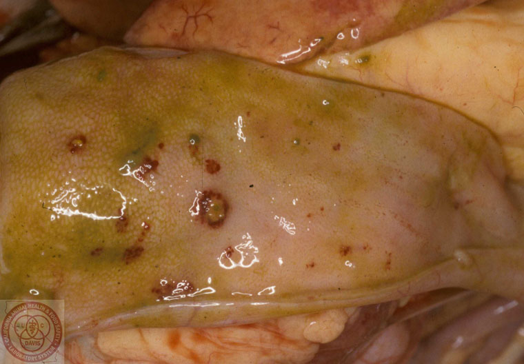
Description:
Avian, proventriculus. The proximal mucosa is eroded and covered by a fibrinonecrotic (diphtheritic) membrane.
Photo ID: ND_013

Description:
Avian, cecal tonsil necrosis.
Photo ID: ND_014
Description:
Avian, diphtheritic laryngo-tracheitis
Photo ID: ND_015
Description:
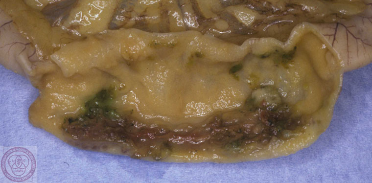
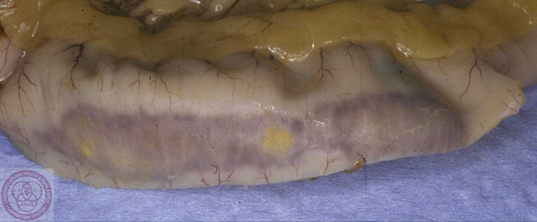
Avian, segmental intestinal necrosis, mucosal.
Photo ID: ND_016
Description:
Avian, segmental intestinal necrosis, mucosal.
Photo ID: ND_017