Disease Images
Disease Images
Disease Images: Epizootic Hemorrhagic Disease
Additional resources for Epizootic Hemorrhagic Disease
Description:
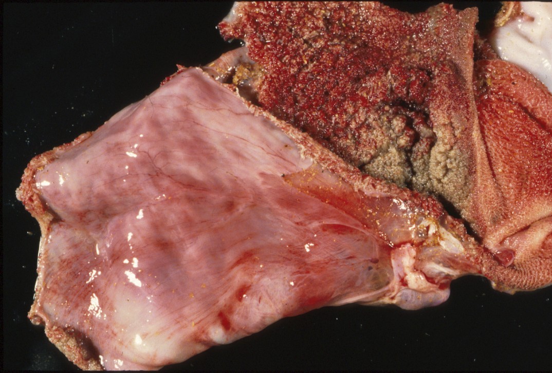
Deer, rumen and reticulum. The serosal surface of the rumen has fine linear to coalescing hemorrhages, and there is extensive congestion and hemorrhage of the ruminal and reticular mucosa.
Photo ID: EHD_001
Description:
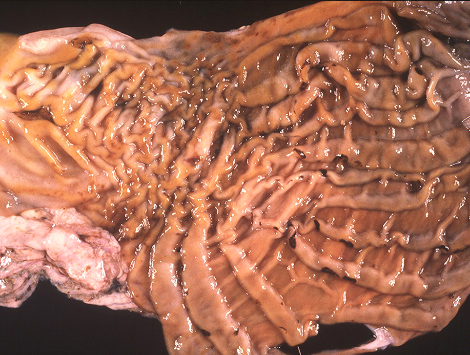
Deer, abomasum. Mucosal folds are diffusely thickened by edema and contain multifocal, sharply-demarcated, red-brown areas of ulceration and hemorrhage.
Photo ID: EHD_002
Description:
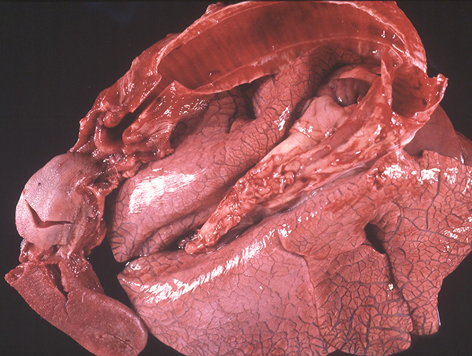
Deer, lungs and trachea. There is moderate to marked widening of interlobular septa (edema). The tracheal mucosa is diffusely congested and contains several blood clots, and there are a few small pleural hemorrhages.
Photo ID: EHD_003
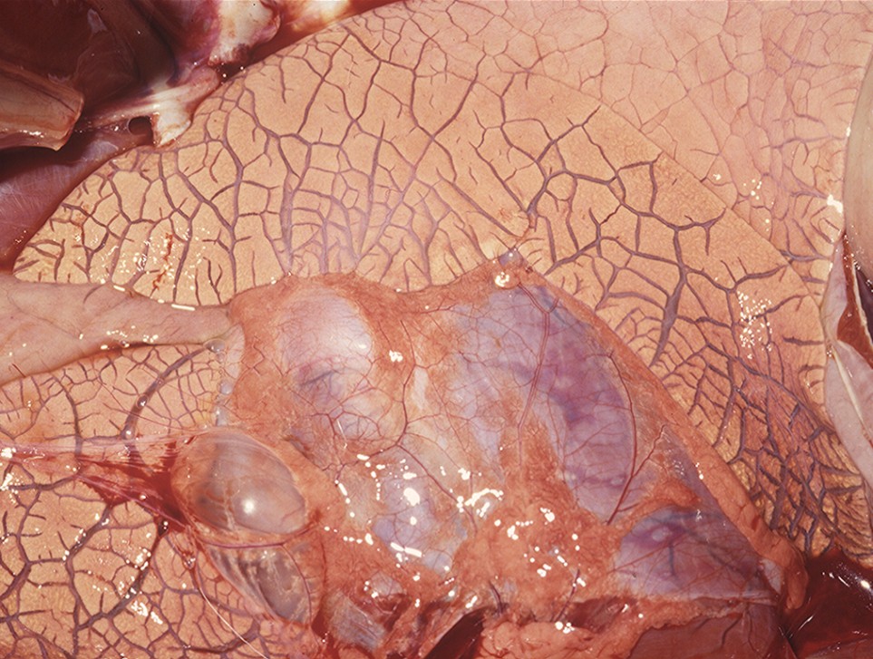
Description:
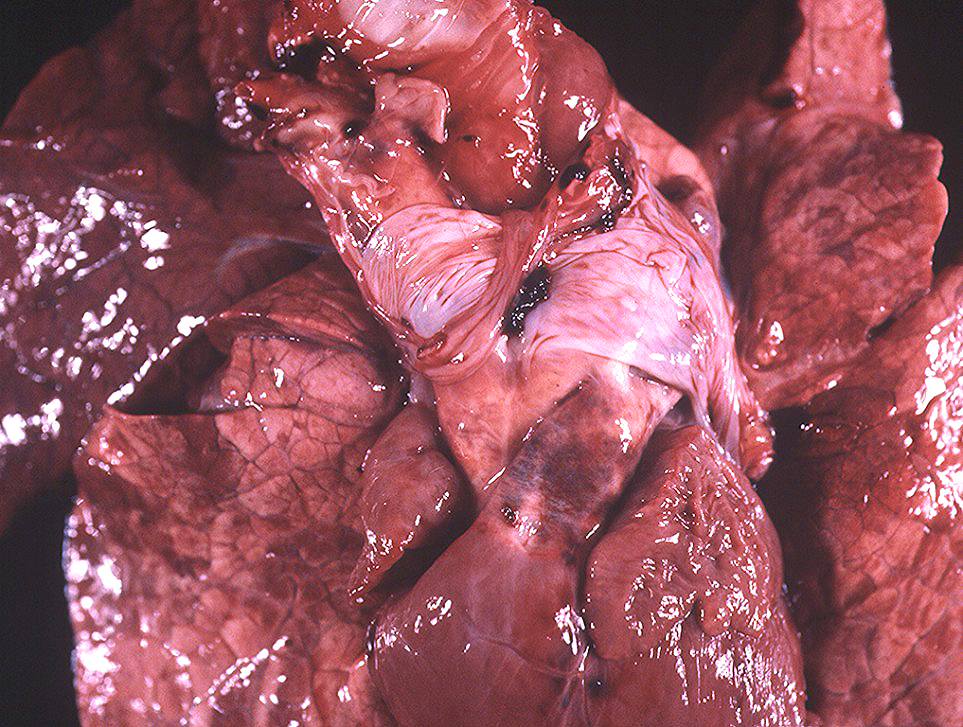
Deer, pulmonary artery and lungs. Locally extensive adventitial hemorrhage at the base of the pulmonary artery is considered pathognomonic for EHD and bluetongue in ruminants. Lungs contain many red areas (congestion and hemorrhage).
Photo ID: EHD_004
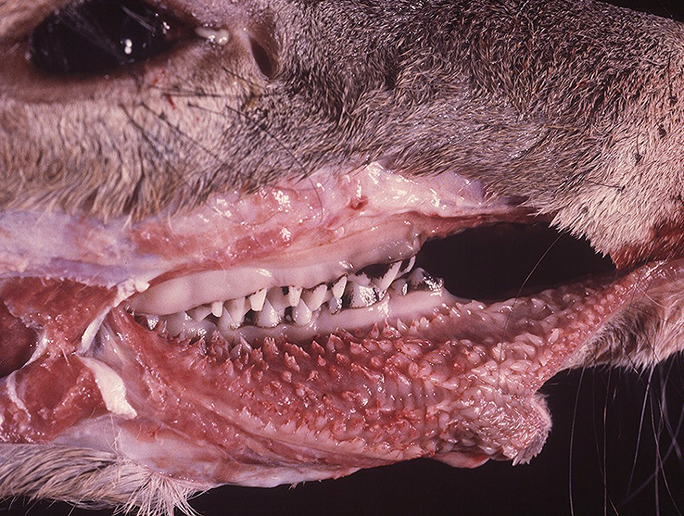
Description:
Deer, oral mucosa. The tips of many buccal papillae are reddened and eroded.
Photo ID: EHD_005
Description:
Deer, lungs. Interlobular septa are severely expanded by edema. The mediastinal bulla likely resulted from terminal dyspnea.
Photo ID: EHD_006

Description:
Deer, rumen and reticulum (preserved specimen). The mucosa contains numerous, variably-sized areas of erosion and ulceration.
Photo ID: EHD_007