Disease Images
Disease Images
Disease Images: Bovine Tuberculosis
Additional resources for Bovine Tuberculosis
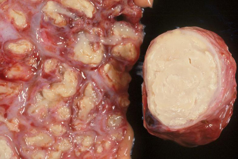
Description:
Elk, lung & lymph node. The lung contains multiple coalescing foci of caseous necrosis surrounded by thin pale fibrous tissue capsules (tubercles). Most of the lymph node is replaced by caseonecrotic debris with a laminated appearance reminiscent of caseous lymphadenitis caused by Corynebacterium pseudotuberculosis.
Photo ID: TUB_001
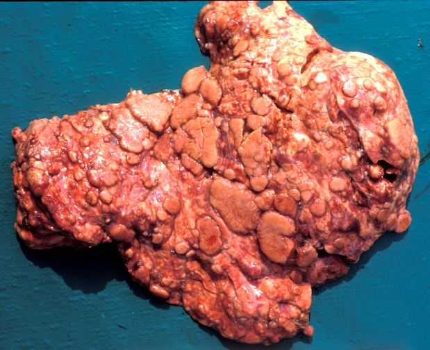
Description:
Bovine, lung. Lung parenchyma is almost entirely replaced by variably-sized, coalescing, raised pale nodules.
Photo ID: TUB_002
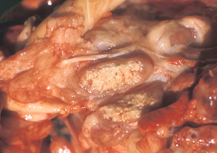
Description:
Pig, tracheobronchial lymph nodes. The center of the sectioned node is replaced by caseous, mineralized debris.
Photo ID: TUB_003
Description:
Bovine, uterus. The endometrium contains numerous raised tubercles.
Photo ID: TUB_004
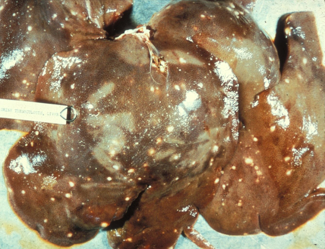
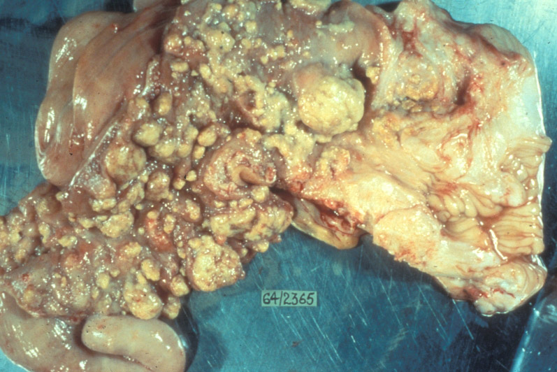
Description:
Pig, liver. Pale, slightly raised granulomas are disseminated throughout all liver lobes.
Photo ID: TUB_005
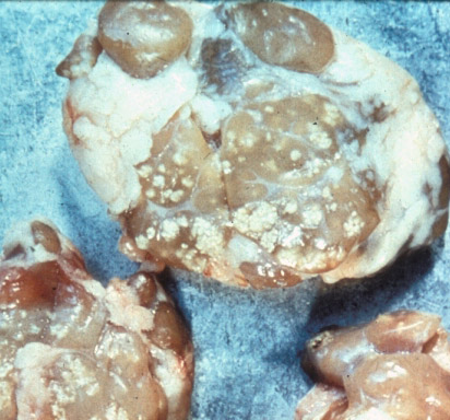
Description:
Pig, lymph node. Pale, mineralized granulomas are scattered throughout these cervical lymph nodes.
Photo ID: TUB_006