Disease Images
Disease Images
Disease Images: Contagious Bovine Pleuropneumonia
Additional resources for Contagious Bovine Pleuropneumonia
Description:
Bovine, lungs. Most of the pleural surface is covered by abundant fibrin and fibrous tissue.
Photo ID: CBP_001

Description:
Bovine, pleural cavity. Large sheets of fibrin cover the costal and diaphragmatic pleura, and form pockets containing straw-colored fluid.
Photo ID: CBP_002
Description:
Bovine, pleural cavity. There is a thick plaque (adhesion) of fibrous tissue on the costal pleura.
Photo ID: CBP_003
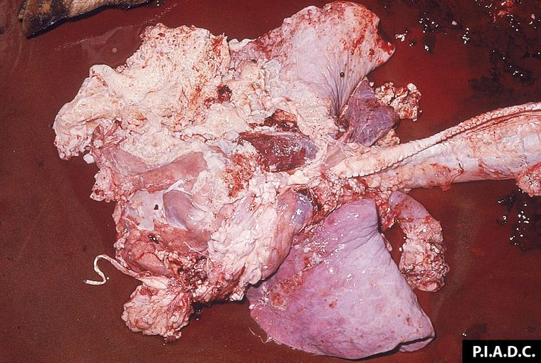
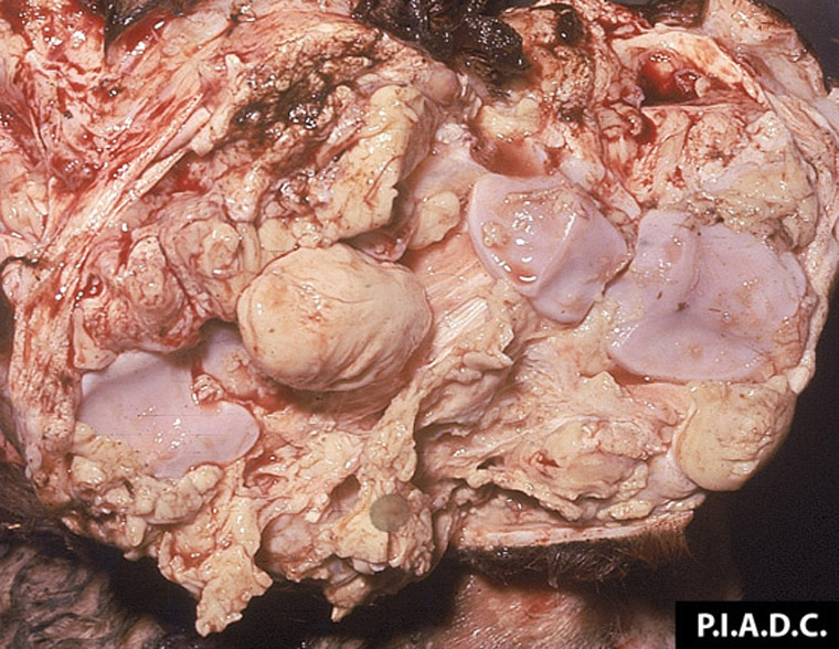
Description:
Bovine, lung. Most of the parenchyma is dull, tan, and contains multiple cavities (necrotic); since it is partially surrounded by a fibrous capsule, this necrotic zone is termed a sequestrum. In the viable tissue above and below the sequestrum, the interlobular septa are markedly thickened by fibrous tissue.
Photo ID: CBP_004
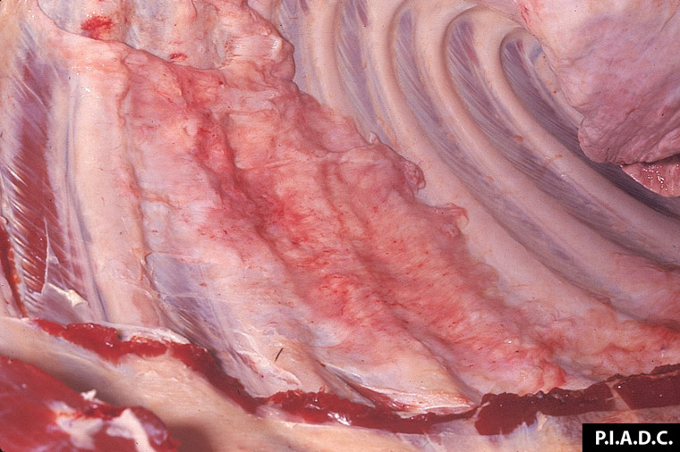
Description:
Bovine, lung. Interlobular septa are markedly thickened by fibrous tissue, and also contain small depressions (air pockets = emhysema). Lobules are reddened and wet (congestion and edema).
Photo ID: CBP_005

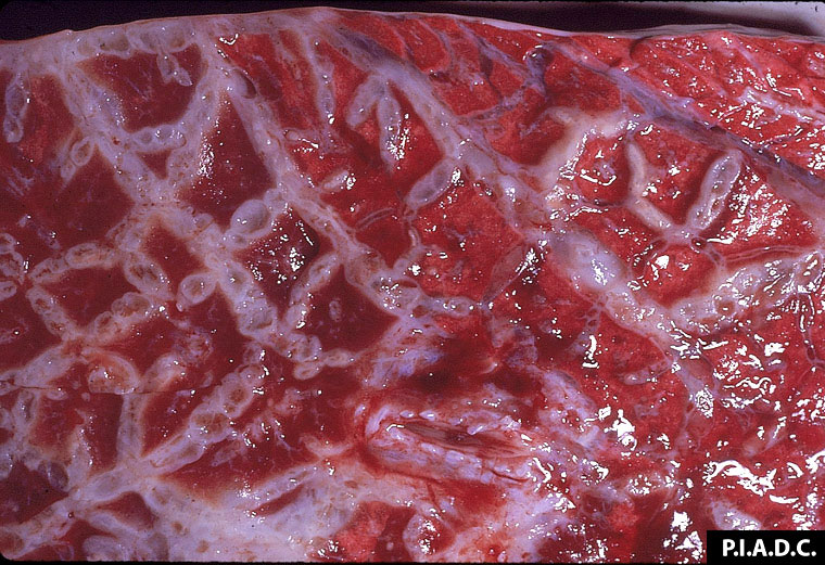
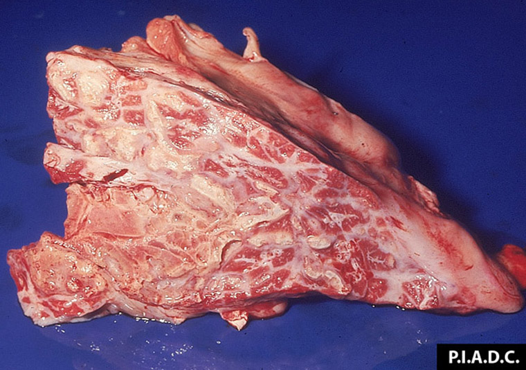
Description:
Bovine, lung. In the ventral portion of this lung (left side of the image), interlobular septa and the pleura are markedly thickened with fibrous tissue; this pneumonic lung is sharply demarcated from the relatively normal dorsal portion tissue.
Photo ID: CBP_006
Description:
Bovine, lung. The pleura and underlying interlobular septa are severely thickened by fibrous tissue. Lung parenchyma at the lower left is dull and tan (sequestrum).
Photo ID: CBP_007

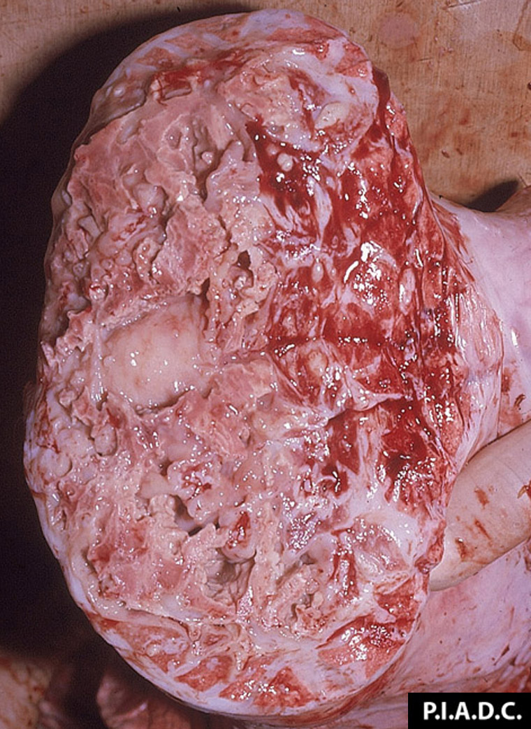
Description:
Bovine, tracheobronchial lymph node. This bisected node is enlarged (hyperplasia) and contains a focal area of hemorrhage.
Photo ID: CBP_008
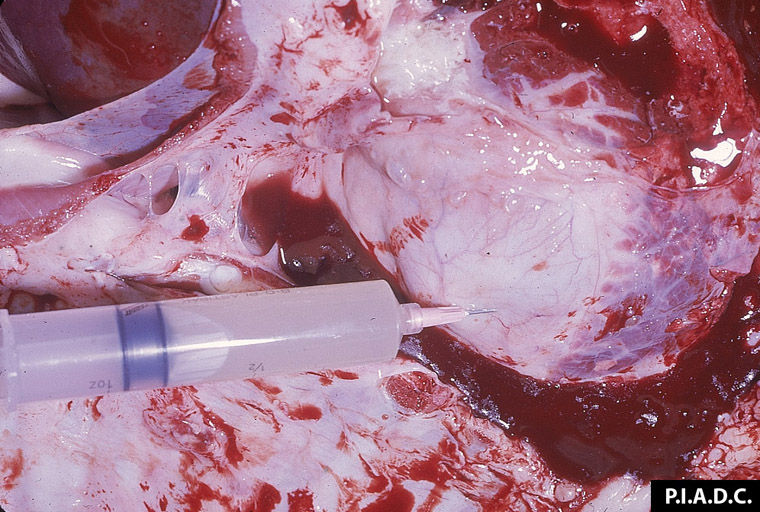
Description:
Bovine, heart. The pericardial sac contains abundant pale turbid fluid.
Photo ID: CBP_009

Description:
Bovine, heart. The pericardial wall is markedly thickened, and the pericardial sac contains abundant pale tan, turbid fluid.
Photo ID: CBP_010
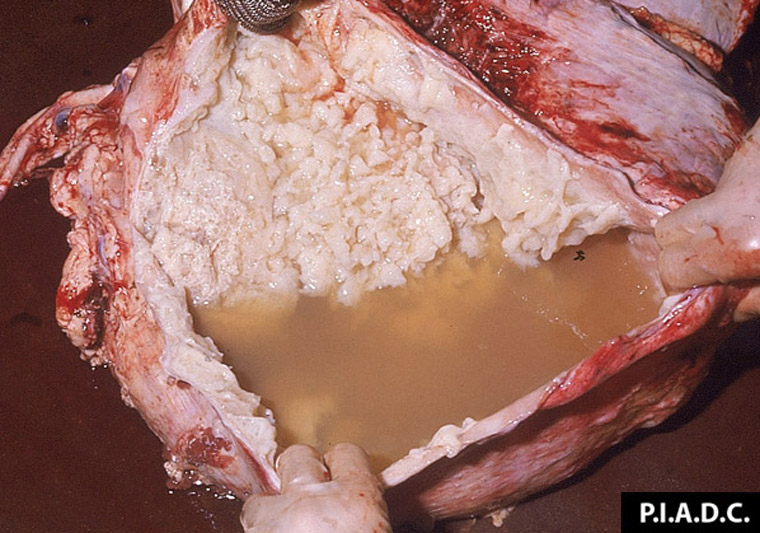
Description:
Bovine, incised pericardium. The sac is distended with abundant turbid, tan fluid, and abundant fibrin coats the pericardial surfaces.
Photo ID: CBP_011
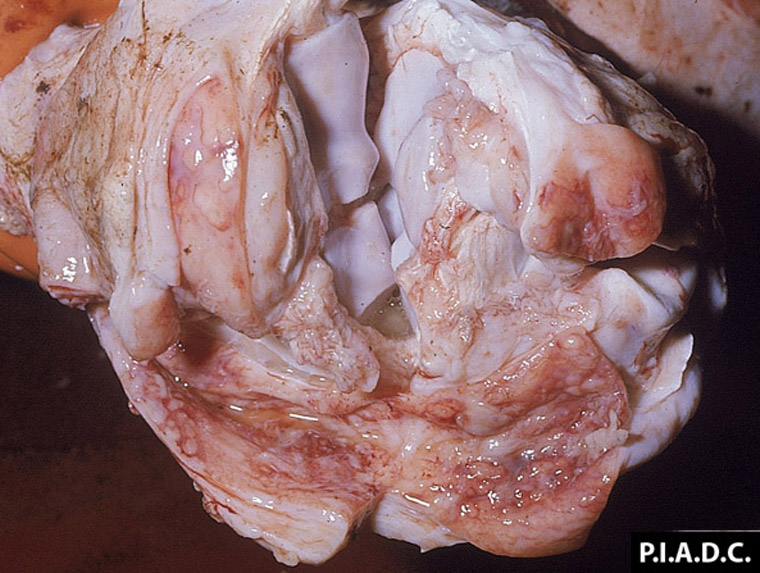
Description:
Bovine, carpus. The joint capsule and adjacent extensor tendon sheath are markedly thickened and contain excessive fluid. The tendon sheath synovium is congested and covered by small flecks of fibrin.
Photo ID: CBP_012
Description:
Bovine, carpus. There is abundant fibrin within the synovial space and on the synovium, and articular cartilages contain a few small erosions.
Photo ID: CBP_013