Disease Images
Disease Images
Disease Images: Q Fever
Additional resources for Q Fever
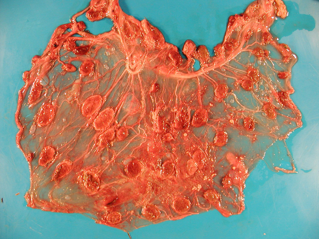
Description:
Goat, placenta. Most cotyledons have pale tan margins (necrosis). There are scattered raised tan discrete intercotyledonary plaques (exudate) and a locally extensive area where the intercotyledonary placenta is opaque, thickened and tan.
Photo ID: QF_001
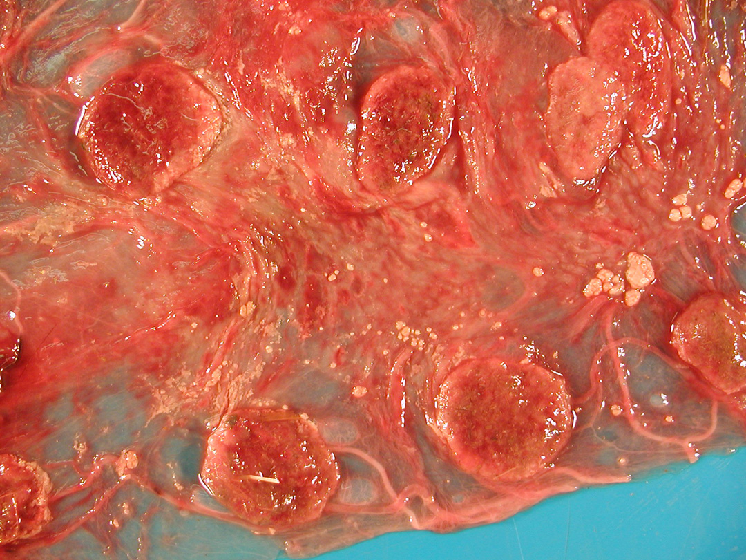
Description:
Goat, placenta. The intercotyledonary placenta is thickened, opaque, and multifocally covered by tan clumps of exudate. Margins of several cotyledons are tan (necrosis), and centers are mottled red-brown (congestion and exudation).
Photo ID: QF_002