Disease Images
Disease Images
Disease Images: Akabane
Additional resources for Akabane
Description:
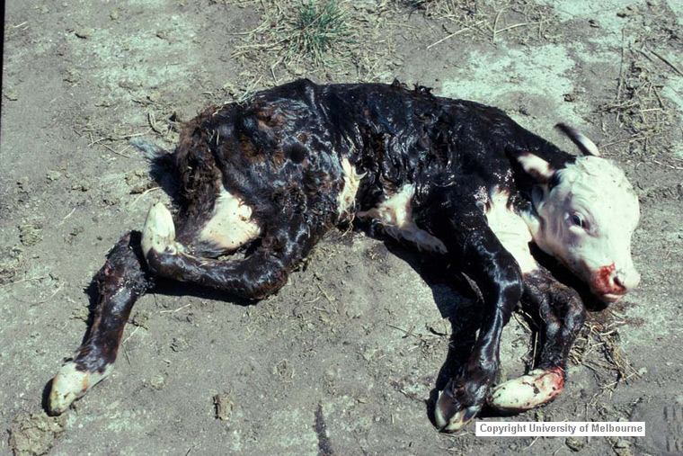
Bovine neonate. This live calf cannot stand due to severe arthrogryposis, primarily affecting the hindlimbs.
Photo ID: AKA_001
Description:
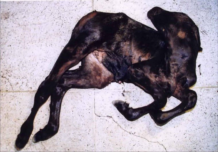
Bovine neonate (Aino). This stillborn calf exhibits torticollis and arthrogryposis.
Photo ID: AKA_002
Description:
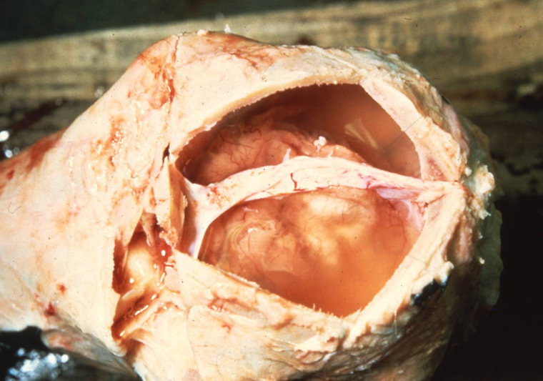
Bovine neonate, brain. The entire brain is reduced in size (microencephaly), and surrounded by cerebrospinal fluid.
Photo ID: AKA_003
Description:
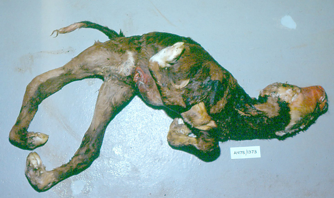
Bovine calf. The head is hyperextended. The limb joints are fixed and vary from hypercontracted to hyperextended.
Photo ID: AKA_004
Description:
Bovine calf, calvarium. The cerebral hemispheres consist of thin-walled sacs that contained cerebralspinal fluid prior to necropsy.
Photo ID: AKA_005
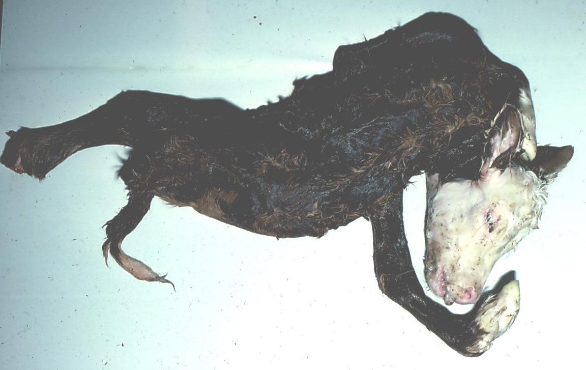
Description:
Bovine calf. The head is rotated and tilted to the side (torticollis). There is abnormal rotation of the thoracic limbs and the joints are fixed at unusual angles (arthrogryposis). The thoracolumbar spine is curved to the right (kyphosis).
Photo ID: AKA-006

Description:
The cerebral hemispheres have failed to develop.
Photo ID: AKA_007

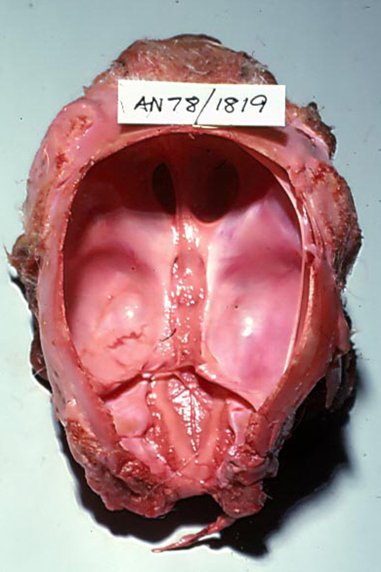
Description:
Bovine steer, calvarium. The cerebral hemispheres are moderately to severely reduced in size and do not fill the cranial vault; the resulting potential space contained cerebral spinal fluid (external hydrocephalus/ hydranencephaly).
Photo ID: AKA_008